Download
1 / 16
160 likes | 326 Views
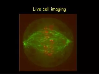
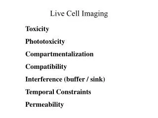
Live Imaging and Other Capabilities of the Bioscience Imaging Facility (BIF). The Bioscience Imaging Facility. The BIF provides the instruments and expertise needed to visualize molecules in preparations ranging from single cells to entire animals.

E N D
Live Imaging and Other Capabilities of the Bioscience Imaging Facility (BIF)
The Bioscience Imaging Facility The BIF provides the instruments and expertise needed to visualize molecules in preparations ranging from single cells to entire animals. • Project specific instrument training & advice • Consultation on sample preparation, image processing, and data analysis • Charge-back rates; Collaborations are encouraged
Confocal Microscopy Nikon A1R • High NA oil objectives 10x, 20x, 40x, 60x • Seven visible laser lines 446, 458, 488, 514, 561, 640 nm • Four channels or 32-bin spectral detection • Resonant scanner* • Piezo stage* • Live imaging chamber* *Ideal for live imaging monolayer cell cultures • High NA oil objectives 10x, 20x, 40x, 60x • Seven visible laser lines 446, 458, 488, 514, 561, 640 nm • Four channels or 32-bin spectral detection • Resonant scanner & Piezo stage* • Live imaging chamber* *Ideal for live imaging monolayer cell cultures Nikon C1+ • High NA oil objectives 10x, 20x, 40x, 60x • Seven visible laser lines 446, 458, 488, 514, 561, 640 nm • Four channel detection • High NA oil objectives 10x, 20x, 40x, 60x • Seven visible laser lines 405, 458, 488, 514, 561, 640 nm • Four channel detection Rotate and Adj
High Speed Image Acquisition Piezoelectric z-stage Resonant Galvanometer
Spectral Acquisition Spectral Unmixing Spectral Detector T(λ) = C1•R1(λ) + C2•R2(λ) Other Uses: • -Document spectra • -Remove autofluorescence • --Spectral FRET • --Create virtual channels Fluorophores in sample
Multiphoton Microscopy Nikon A1R_MP • LWD (500 um) water 40x objective* • Mai Tai DeepSee tunable IR laser* • Four channel non-descanned detection* • Resonant scanner* • Live imaging chamber* • Confocal option Two laser lines (405, 488); four channel detection. *Ideal for live imaging deep into living tissues
Wide-field Microscopy Nikon Ti-S • • Phase and PlanFluor objectives* • • DS-Ri1 color camera (standard) • • PhotometricsQuantEM CCD (optional)* • • Live imaging chamber* • *Ideal for high sensitivity or high frame rate imaging of monolayer cell cultures
Microscopic Images Pablo Zavattieri Civil Engineering Juan Martinez, Mark Hall Biochemistry Aaron Taylor Bindley Shawn Liu, Xiaoqi Lin Biology
In vivo Optical Imaging IVIS Lumina II • • Cooled CCD • • Range of excitation and emission filters • Spans the 450-770 nm • • Isofluorane anesthesia provided • *Ideal for collecting signal from within an organism
In vivo Optical Images Dan Szymanski Agronomy CansuCimen, Tim Ratliff Comparative Pathobiology
Micro SPECT / CT Imaging MiLabs U-SPECT-II/CT • • Combined SPECT and x-ray CT • Enables SPECT and CT image registration • • Multi-pinhole collimators • High resolution imaging (350 um voxels) • High sensitivity imaging (kBeq source) • • Data binning relative to biological rhythms • Reduces blur • • List mode acquisition and statistical 3-D • image reconstruction algorithm. • Improves image quality
Principles of Statistical Reconstruction Object Space ? Projection Space
Micro SPECT / CT Images Charity Wayua, Phil Low Chemistry Xin Lu, Shuang Liu Health Sciences
Thank You! For more information: Email:abtaylor@purdue.edu Web:http://www.purdue.edu/discoverypark/bioscience/facilities/imaging/index.php