Download
1 / 42
420 likes | 695 Views
IRON DEFICIENCY AND RELATED HYPOCHROMIC ANEMIAS. Problems with heme synthesis. IRON DEFICIENCY AND RELATED ANEMIAS. Iron, as previously discussed, is crucial to O 2 transport. A disorder in iron metabolism may result in iron deficiency anemia. Normal iron metabolism:

E N D
IRON DEFICIENCY AND RELATED HYPOCHROMIC ANEMIAS Problems with heme synthesis
IRON DEFICIENCY AND RELATED ANEMIAS • Iron, as previously discussed, is crucial to O2 transport. A disorder in iron metabolism may result in iron deficiency anemia. • Normal iron metabolism: • Iron is the most abundant trace element in the body • 2/3 of total body iron is in heme
IRON DEFICIENCY AND RELATED ANEMIAS • Each day 20-25 mg of iron is needed for erythropoiesis, most of which is salvaged from normal RBC turnover and hemoglobin catabolism. • In an adult male, only about 1 mg/day needs to be newly absorbed from the diet. • Menstruating females need to absorb more per day because 30-40 mg is lost per month. Therefore, they need to absorb an additional 1-1.5 mg/day on top of the male requirement of 1 mg/day • There is an increased need for iron during • Pregnancy • Childhood • Adolescence
IRON DEFICIENCY AND RELATED ANEMIAS • 1/3 of the body’s total iron is stored in the liver, spleen, or bone marrow, or is carried in myoglobin and coenzymes of the cytochrome electron transport chain. • Iron containing proteins are classified as: • Heme – contains a porphyrin-iron complex. Includes • Hemoglobin • Myoglobin • Catalases • Peroxidases • Cytochrome electron transport proteins
IRON DEFICIENCY AND RELATED ANEMIAS • Non-heme proteins – includes • A large number of sulfur iron-binding compounds • The iron transport proteins • Transferrin • Lactoferrin • Iron storage proteins • Ferritin (for short term storage) • Hemosiderin (for long term storage) • Iron absorption – • Is regulated by a natural intestinal mechanism in which G.I. mucosal cells admit just enough iron to cover losses from normal metabolic turnover of RBCs
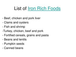
IRON DEFICIENCY AND RELATED ANEMIAS • Maximum iron absorption occurs in the duodenum and upper jejunum • An increased motility of nutrients through the G.I. tract or a decreased absorptive surface area in the G.I. tract may result in decreased iron absorption. • The amount of iron absorbed depends upon: • The amount and type of iron ingested (Non-heme iron, which is in the ferric form, in vegetables and whole grains is not easily absorbed, while the heme iron, which is in the ferrous form and present in red meats, is more readily absorbed) • The presence or absence of other food (other food may interfere with the uptake of iron) • G.I. acidity (an acidic condition favors uptake)
IRON DEFICIENCY AND RELATED ANEMIAS • State of iron stores in the body (less is absorbed if iron stores are high) • Activity of the bone marrow (more iron is absorbed when erythropoietic activity is increased) • State of the intestinal mucosa • In a state of severe iron deficiency, the body can increase the amount of iron absorbed up to 30% of the dietary iron (normally only 10% is absorbed) to compensate for the iron depletion • Iron transport and storage • Iron is transported from the duodenum into mucosal cells in the ferrous state where it is converted to the ferric state.
IRON DEFICIENCY AND RELATED ANEMIAS • It may combine with apoferritin to form ferritin or cross into the plasma • In the plasma two atoms bind to the iron transport protein, transferrin. • The total iron binding capacity of the serum (TIBC) is a functional measurement of the amount of transferrin. • Normally the transferrin in the serum is about 30% saturated with iron. • The majority of the transferrin bound iron is delivered to the bone marrow where it binds to specific transferrin receptors on the developing RBCs (normoblasts) and releases the iron in the ferrous state.
IRON DEFICIENCY AND RELATED ANEMIAS • Iron in excess of the erythropoietic requirements is delivered to the liver and other tissues where it unloads the iron which is then stored bound to : • Apoferritin as ferritin • The amount of apoferritin produced is dependent upon the storage needs and iron available from the diet. • In iron deficiency, little is made and low levels of ferritin in the serum (although primarily intracellular, small amounts are always entering the blood) may be the first sign of a developing iron deficiency anemia • When iron is absorbed in excess of the ferritin storage capacity, denatured ferritin and excess iron are deposited as hemosiderin (seen in about 25% of developing RBCs in the bone marrow.
IRON DEFICIENCY AND RELATED ANEMIAS • When the body needs iron immediately, iron is released from tissue ferritin and transported via transferrin, to developing erythrocytes in the bone marrow • As a general rule: Know this! • As serum iron increases, storage iron (ferritin) increases and transferrin and transferrin receptors decrease. • As serum iron decreases, storage iron (ferritin) decreases, and transferrin and transferrin receptors increase • The way to increase absorption of iron from the GI tract is to increase the production of transferrin (by liver) and transferrin receptors which bind iron saturated luminal transferrin
HOW IRON LEVELS AFFECT FERRITIN AND TRANSFERRIN RECEPTOR SYNTHESIS IRE-BP Sufficient cellular iron Depleted cellular iron IRE-BP has low affinity for RNA binding IRE-BP has a high affinity for RNA binding Translation of ferritinmRNA Decreased stability of transferrin receptor mRNA Translation of ALAS mRNA Inhibits translation of ferritin RNA Stabilizes transferrin receptor mRNA Inhibits translation of ALAS Increased ferritin Decreased transferrin receptors Heme synthesis stimulated Decreased ferritin Increased transferrin receptors Heme synthesis decreased
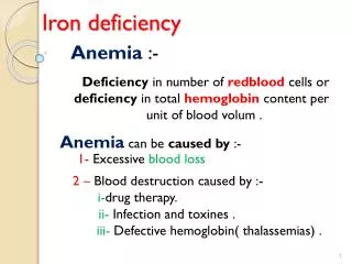
CLINICAL SYNDROMES • Iron deficiency anemia (IDA) – is also called sideropenic anemia • Is the most common nutritional deficiency in the world • Etiology of a state where body iron stores are depleted • Poor dietary supplementation is actually rare because iron is found in many foods – deficiency occurs most commonly where grain is the mainstay of the diet • Diminished absorption of iron – celiac disease, defective gastric function, achlorhydria, following gastrectomy • Distribution defects – rare congenital deficiency of transferrin
CLINICAL SYNDROMES • Increased requirements such as in pregnancy, during infancy or adolescence • Excessive blood loss – is most commonly found in menstruating women. • May also be found with GI blood loss which may occur in peptic ulcers, diverticulosis, inflammatory bowel disease, hemorrhoids, hookworm infections, certain malignancies. • Chronic intravascular hemolysis can also lead to IDA. • The anemia: • Symptoms include fatigue, lethargy, and dizziness • Signs include • pallor of mucous membranes, • bounding pulse, • systolic flow murmurs, • smooth tongue, • koilonychias (spooning of nails), and
ESOPHAGEAL WEBS • esophageal webs (manifested as "shelves" of tissue extending into the esophagus that may cause dysphagia - difficulty in swallowing) http://img.medscape.com/pi/emed/ckb/gastroenterology/169972-186561-2487.jpg
CLINICAL SYNDROMES • Hematologic indices – hypochromic, microcytic with moderate to severe anemia (more on this later) • The deficiency occurs in three phases • Iron depletion – ferritin and hemosiderin stores are depleted, iron absorption and TIBC begin to increase. • Iron deficient erythropoiesis where the RBC continues to make porphyrin even in the absence of iron – serum iron levels decrease, TIBC increases, and the % saturation decreases, transferrin receptors on red cells increase • Iron deficiency anemia – hgb, hct, and MCV all decrease • The anemia occurs in three stages
CLINICAL SYNDROMES • Normochromic, normocytic with anisocytosis • Microcytic with anisocytosis • Hypochromic, microcytic– in severe cases, poikilocytosis may be marked and nucleated RBCs may be seen in a peripheral smear • Free erythrocyte protoporphyrin increases • The reticulocyte count is decreased in relation to the severity of the disease • Decreased serum ferritin levels • In the bone marrow sideroblasts are absent or reduced and there is mild to moderate erythroid hyperplasia without increased reticulocytes. • This is really a type of ineffective erythropoiesis due to a decreased ability of the RBC to make hemoglobin within the RBC
CLINICAL SYNDROMES • A defect in iron supply as well as inadequate protoporphyrin synthesis or inadequate globin synthesis can result in a hypochromic, microcytic anemia. • The decreased RBC size is due to a failure of hemoglobin formation to signal to the RBC to stop dividing – there is a nuclear to cytoplasmic maturation deficiency. The cytoplasmic maturation lags behind the nuclear maturation and the cell keeps dividing, getting smaller with each division
CLINICAL SYNDROMES • Other clinical consequences of iron deficiency include: • The iron containing enzyme myeloperoxidase is decreased – this functions in phagocytosis and bacterial killing • There is decreased cell mediated immunity (this means that there is a decreased response to antigenic stimulation) • Splenomegaly may occur in severe cases • Headaches and irritability are common • Early muscle fatigue • Pica – a perversion of appetite
CLINICAL SYNDROMES • The tongue and oral lesions are reversible • The esophageal webs are irreversible • Gastric mucosal atrophy may occur • Therapy • Treat the underlying disorder (for example, correct the cause of bleeding) • Treat the iron deficiency usually by oral administration of ferrous sulfate. • With absorption problems, parenteral administration may be necessary. • The response will occur in three stages: • 3-5 days – there will be an improvement in the feeling of well being
CLINICAL SYNDROMES • 7-10 days – the reticulocyte count will increase and in 6-10 weeks the hemoglobin level should be normal • Replacement of iron storage supplies will occur over a period of months • Iron overload • Hemosiderosis is an accumulation of iron to supranormal levels with deposition of iron primarily in the macrophages of the spleen, liver, bone marrow, and other tissues.
CLINICAL SYNDROMES • Causes of hemosiderosis are • Hemolytic anemias • Multiple blood transfusions • Disorders of erythropoiesis • Inappropriate intestinal absorption of iron • There is usually no organ damage • Hemochromatosis occurs when iron accumulation has progressed to involve widespread deposition of iron in parenchymal tissue with resulting organ injury. • Excess iron deposits in hepatocytes, cardiac cells, endocrine cells, and other parenchymal cells can interfere with the their normal function and may lead to death.
CLINICAL SYNDROMES • Signs include • Skin pigmentation because of an effect on melanin synthesis • Diabetes mellitis • Hepatomegaly with or without heart disease and hypogonadism. • Idiopathic hemochromatosis is an inherited abnormality with defective regulation of iron absorption leading to high levels of iron deposition in parenchymal tissues • Serum ferritin and iron levels are increased
CLINICAL SYNDROMES • The TIBC is normal (if everything were working correctly, what should it be?) • The transferrin is 100% saturated • Treatment for hemachromatosis is phlebotomy and/or infusion of iron chelators to bind and excrete the iron. • Disorders of iron metabolism – can result from a block in incorporation of iron or there may be defective iron utilization. Either of these will lead to a hypochromic, microcytic anemia.
CLINICAL SYNDROMES • Sideroblastic anemias – are characterized by abnormalities of heme metabolism with increased numbers of ringed sideroblasts in the bone marrow and an increase in total body iron. There are two major groups of sideroblastic anemias: • Inherited – may be sex-linked or recessive – both are rare • Defective heme synthesis is involved in the pathogenesis of this. • The most common defect is sex-linked and is due to an abnormal ALA synthase enzyme (involved in the first step in heme synthesis) as a result of heterogenous point mutations in the catalytic domain of the enzyme.
CLINICAL SYNDROMES • The activity of ALA synthase is totally dependent upon the presence of the cofactor pyridoxal phosphate. The coenzyme binds to the enzyme and is crucial for its stability, and its catalytic activity. With some of the mutant variants of the enzyme, administration of pyridoxal phosphate may provide a partial to complete resolution of the disease. • Decreased heme synthesis due to a block in iron utilization is interpreted by the body as an increased need for iron. • Therefore, there will be increased absorption of iron which will then accumulate in the tissues (results in the increased ringed sideroblasts in the bone marrow). • Acquired • Primary idiopathic = refractory anemia with ringed sideroblasts (RARS) – there appear to be multiple mitochondrial enzyme defects including a decrease in ALA synthase activity. • May terminate in an acute leukemia or other malignant disorder.
CLINICAL SYNDROMES • Secondary to drugs or toxins that interfere with the activity of heme synthesis. This includes: • Chemotherapeutic drugs, antituberculosis drugs, or chloramphenicol • Lead poisoning – lead may affect many of the enzymatic steps in heme synthesis. • The most dramatic effects are on the conversion of ALA to prophobilinogen and on the incorporation of iron into protoporphyrin IX. • This leads to increased urine excretion of ALA, increased erythrocyte protoporphyrin, and iron accumulation in cells • Chronic alcohol intoxication – can lead to several different kinds of anemia including a sideroblastic anemia because alcohol inhibits the synthesis of pyridoxal phosphate, the cofactor required for ALA synthase activity.
CLINICAL SYNDROMES • Associated with a malignancy such as leukemia, multiple myeloma, or lymphoma. • Clinical and lab features in sideroblasic anemias include: • In anemias secondary to drugs or malignancy, manifestations of the primary disorder are dominant • In primary, idiopathic anemia (RARS) hepato and splenomegaly are common, as is diabetes. • Cardiac problems occur as the disease progresses • A study of the peripheral blood reveals: • Two RBC populations (dimorphism) normochromic, normocytic (or macrocytic) and hypochromic, microcytic which is a reflection of the disturbed iron utilization
CLINICAL SYNDROMES • MCV, MCHC, and MCH may be normal (particularly with macrocytic and microcytic cells) • RDW is increased (increased anisocytosis) • Poikilocytosis may be present • RBC may contain pappenheimer bodies (iron granules) • Basophilic stippling may be seen – in lead poisoning, the basophilic stippling is very coarse • Protoporphyrin levels are usually increased • Serum iron is increased • The TIBC is normal or decreased with increased saturation of transferrin • The bone marrow is hyperplastic with ineffective erythropoiesis occurring and 40% or more ringed sideroblasts.
CLINICAL SYNDROMES • Treatment • Pyridoxine therapy with hereditary forms of the disease (may or may not help) • Primary idiopathic forms are treated with blood transfusions • Secondary forms – treat the underlying disease or eliminate the drug • Anemia of chronic disorders – can occur with chronic infections, chronic non-infectious inflammatory responses, malignant neoplasms, autoimmune disorders such as SLE, and chronic renal disease. • Three problems occur and are due to cytokines released by the activated immune system:
CLINICAL SYNDROMES • A block in the release of iron from macrophages for recycling. This results in decreased iron flow to the bone marrow • No stimulation of erythropoietin production despite the anemia (cytokines inhibit erythropoietin production) • RBC survival is shortened due to extracorpuscular factors • Clinical and laboratory features include: • Signs and symptoms associated with the underlying disease • Mild anemia that may be normochromic, normocytic, hypochromic, or hypochromic, microcytic (hypo preceeds micro) • Decreased reticulocyte count
CLINICAL SYNDROMES • Decreased plasma iron • Decreased TIBC • Normal to increased serum ferritin • In the bone marrow there is an increased myeloid (WBC) to erythroid (RBC) ratio, decreased sideroblasts, but increased hemosiderin in macrophages • Therapy – treat the underlying disorder • Porphyrias – are a group of inherited disorders characterized by a block in porphyrin synthesis which occurs in most cells although the erythroid cells in the bone marrow and hepatocytes are most active in terms of porphyrin synthesis.
CLINICAL SYNDROMES • The block in synthesis results in an accumulation of porphyrin heme precursors in the tissues with large amounts excreted in the urine and feces. • The excess porphyrins, which fluoresce in the presence of UV light, are deposited in the tissues and cause most of the symptoms and clinical findings: • Photosensitivity due to excess porphyrins in the skin • Abdominal pain – occurs in hepatic form • Neuropathy (motor dysfunction, sensory loss, and mental disturbance) – occurs in hepatic form • Two forms, erythropoietic and hepatic, are expressed depending upon the primary site of defective porphyrin synthesis.
CLINICAL SYNDROMES • Erythropoietic porphyria • Congenital erythropoietic porphyria – is due to a defect in UPG III cosynthase resulting in a build-up of UPG I and CPG I. There may also be hyperactivity of UPG I synthase since adequate amounts of heme are produced. • Excess porphyrin deposits in the RBCs may lead to a hemolytic anemia • Erythropoietic protoporphyria – probably due to a defect in ferrochetolase (step where iron is incorporated into protoporphyrin), resulting in an overproduction of protoporphyrin. • No anemia occurs in this form of the disease. • Hepatocytes are also deficient in ferrochetolase activity.
SUMMARY OF CONDITIONS ASSOCIATED WITH ABNORMAL HEME SYNTHESIS