Download
1 / 42
420 likes | 443 Views
Delve into the structures and functions of biomacromolecules like carbohydrates, lipids, proteins, and nucleic acids. Learn how these molecules power cells and provide essential energy. 8 Relevant

E N D
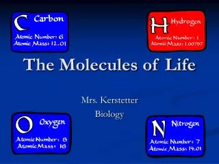
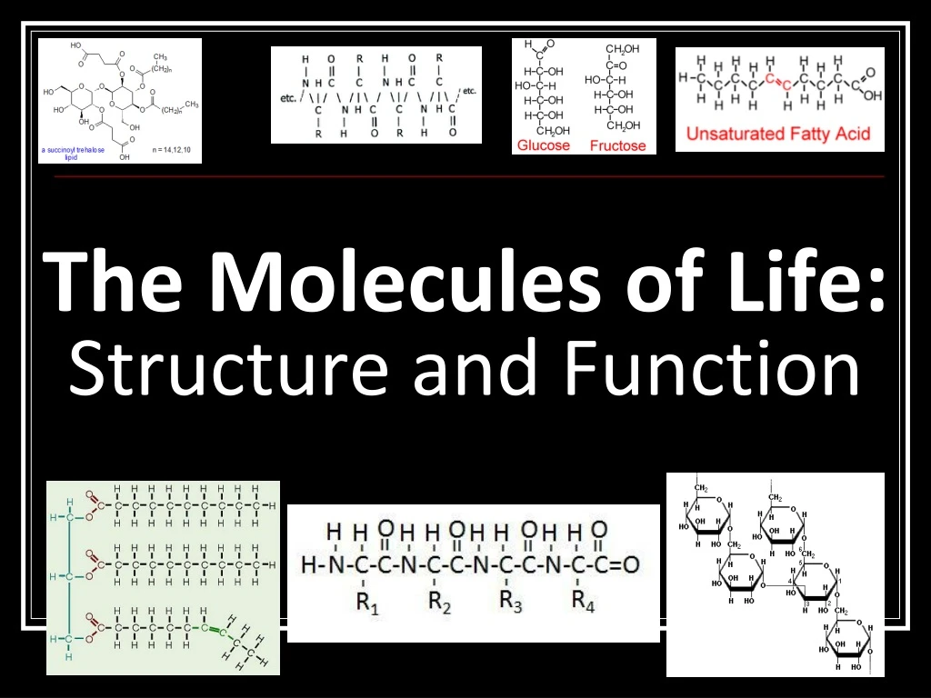
Objective To understand the structure and function of biomac- romolecules and to be able to identify them based on their characteristics. Essential Question: What are the molecules of life, what are their general structures, and functions?
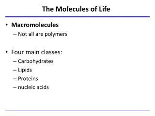
Molecules of Life • “Biomacromolecules” • Carbohydrates • Lipids • Proteins • Nucleic Acids • These make up cells and can be used by cells for energy
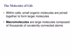
Polymers vs. Monomers Poly = many; Mer = part In biology, a polymer is a large molecule consisting of many smaller sub-units (often repeated) bonded together. Mono = one A monomer is a sub-unit (single unit) of a polymer.
Making a Polymer Dehydration Synthesis reactions Monomers bond to one another through the removal of water. Why? Stores energy Conserves space
Breaking a Polymer Hydrolysis Reactions hydro = water lysis = to break or lyse Polymers are broken down into monomers with the use of water. Why? Access energy To build new polymers
Carbohydrates Molecular formula (C H2 O)n Store energy in chemical structure Glucose most common monosaccharide produced by photosynthetic autotrophs Each “carbon” is surrounded by a “hydrate” (water)
Carbohydrates are classified according to the size of their carbon chains, varies from 3 to 7 carbons Triose = 3 carbons Pentose = 5 carbons Hexose = 6 carbons
Disaccharides “Double sugar” consisting of 2 monosaccharides joined by a glycosidic linkage. What reaction forms the glycosidic linkage?
Example Disaccharides Lactose = glucose + galactose Sucrose = glucose + fructose
Polysaccharides Polymers of a few hundred or a few thousand monosaccharides Function as energy storage molecules or for structural support
Example Carbohydrates Starch = a plant storage from of energy, easily hydrolyzed to glucose units. Polysaccharide. Cellulose = a fiber-like structural material; tough and insoluble. Used in plant cell walls. Polysaccharide. Glycogen = a highly branched chain in animals to store energy in muscles and the liver. Polysaccharide. Chitin = used as a structural material in arthropod exoskeleton and fungal cell walls. Polysaccharide. Lactose = found in milk and dairy products. Disaccharide. Glucose = simplest sugar; used by mitochondria in all cells for energy. Feeds brain. Monosaccharide.
Lipids Large molecules Diverse in structure Nonpolar, so insoluble in water Store energy in chemical structure Groups: Fats, oils, phospholipids, sterols, waxes
Structure of Fatty Acids Long chains of mostly carbon and hydrogen atoms with an acid (-COOH) group at one end Resemble long flexible tails
Saturated vs. Unsaturated Fats Unsaturated fats : liquid at room temp one or more -C=C- (double bonds) between carbons causing “kinks” in the tails most plant fats Saturated fats: solid at room temp only single C-C bonds in fatty acid tails Carbons fully surrounded (“saturated”) with H’s most animal fats
Structure of Triglycerides 1 glycerol + 3 fatty acids Fatty acids and glycerol bound together by ester bonds. Found in food (oils and fats); long term energy storage
Structure of Phospholipids 1 glycerol + 2 fatty acids + phosphate group. Connected by “phosphodiester” bond Main structural component of cell membranes, where they arrange in bilayers.
Waxes Lipids that serve as coatings for plant parts and as animal coverings. Prevents dessication due to insolubility in water.
Steroids Four carbon rings with no fatty acid tails Component of animal cell membranes (cholesterol) Modified to form sex hormones (estrogen, testosterone)
Functions of Lipids Which lipids provide these functions? • Energy storage • Membrane structure • Protecting against desiccation (drying out) • Insulating against cold • Absorbing shock • Regulating cell activities by hormone actions
Proteins 3-dimensional “globular” shape Consist of many peptide bonds between 20 possible amino acid monomers, made by dehydration synthesis Polypeptide = “many” “peptide bond”s; A chain of amino acids
Structure of Amino Acids Amino acids = monomers Consist of an asymmetric carbon bonded to: Hydrogen Amine group Carboxyl (acid) group Variable R group specific to each amino acid
Properties of Amino Acids Grouped by polarity Variable R groups (“side chains”) confer different properties to each amino acid
Example Proteins Enzymes Accelerate specific chemical reactions Structure keratin - found in hair and nails collagen - found in connective tissue Muscle Contraction actin and myosin fibers that interact in muscle tissue
Immune System Function Antibodies recognize and flag foreign substances. Carriers Membrane transport proteins move substances across cell membranes Blood proteins (hemoglobin) carry oxygen throughout the body Signaling and Communication Hormones such as insulin (regulate blood sugar levels) and adrenaline (increase heart rate to adjust to needs) used to help body respond Example Proteins
Recap: Discuss with your group… • Carbohydrate Functions: • Examples include: • How? • Lipid Functions: • Examples include: • How? • Protein Functions: • Examples include: • How?
http://www.youtube.com/watch?v=Oz2x_yxPXww&feature=related&safety_mode=true&persist_safety_mode=1&safe=activehttp://www.youtube.com/watch?v=Oz2x_yxPXww&feature=related&safety_mode=true&persist_safety_mode=1&safe=active • http://www.youtube.com/watch?v=lijQ3a8yUYQ&safety_mode=true&persist_safety_mode=1&safe=active
How to make a Protein in 4 easy steps! Primary Structure Secondary Structure Tertiary Structure Quaternary Structure
Primary Structure Sequence of amino acids in a protein, bonded by peptide bonds This creates the “polypeptide”
Let’s Model the Primary Structure: Salivary Amylase • Observe the properties of the 20 Amino Acids. • What do the different colors represent? • How do you think they would interact?
Make this Primary Sequence: Place amino acids about 1 inch apart (2 finger widths) and fold pieces • White = polar/hydrophilic • Yellow = nonpolar/hydrophobic • Blue = basic (+ charged) • Red = acidic (- charged) • Green = “sulfur R-group” (bonds only Cysteines) MSDKRCTYPCAENQ
Secondary Structure Repeated folding of backbone of polypeptide How? H bonds form between atoms in backbone 2 types: a-helix, b-pleated sheets
Let’s model Secondary Structure • Look at your string of amino acids. • What do the different colors represent? • Note the order of colors. • Take the “backbone” and create some a-helices and some b-pleated sheets.
Tertiary Structure Behavior of R groups determines folding of polypeptide How?Interactions between R groups http://www.youtube.com/watch?NR=1&v=ysPt1lIllcs&safety_mode=true&persist_safety_mode=1&safe=active
Let’s model Tertiary Structure • Note the colors on your polypeptide. • White = polar/hydrophilic • Yellow = nonpolar/hydrophobic • Blue = basic (+ charged) • Red = acidic (- charged) • Green = “sulfur R-group” (bonds only Cysteines)
Quaternary Structure 2 or more polypeptides bonded together How? Attraction between backbones and R groups of neighboring globs
Let’s model Quaternary Structure • Find a neighbor, and attach R groups that might be attracted to each other. What types would?
Factors That May Impact Protein Folding Depends on physical conditions of environment pH, temperature, salinity, etc. Change in environment may lead to denaturation of protein Denatured protein is biologically inactive Can renature if primary structure is not lost What happens when protein folding goes wrong? http://www.youtube.com/watch?NR=1&v=H2Ouxl_GNjA&safety_mode=true&persist_safety_mode=1&safe=active http://www.youtube.com/watch?v=RNIwwLdDLnI&feature=related&safety_mode=true&persist_safety_mode=1&safe=active
Your Tasks • Protein Activity Wrap Up • Stamps for journal activity (models) • Draw your last diagram! Homework due Thursday The Structure and Function of Macromolecules Reading (linked to website) and WS