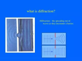
what is diffraction?
what is diffraction?. Diffraction – the spreading out of waves as they encounter a barrier. What is a Diffraction pattern? an interference pattern that results from the superposition of waves.

what is diffraction?
E N D
Presentation Transcript
what is diffraction? Diffraction – the spreading out of waves as they encounter a barrier.
What is a Diffraction pattern? • an interference pattern that results from the superposition of waves. • Mathematically, this process can be described by Fourier transform, if the diffraction is kinematic (electron or X-ray has been scatted only once inside the object). Laser diffraction pattern of a thin grating films, where the size of holes is closed to the wavelength of the laser (Ruby red light 594 um) .
Fourier transform of regular lattices: Real space Reciprocal space
What is a crystal ? DNA single crystal single crystal of quartz • Same structural unit (an atom, many atoms, molecule) – unit cell. • The units are packed periodically in a ‘infinite’ space – lattice. • Unit cell contains all the necessary points on the lattice that can be translated to repeat itself in an infinite array. In other words, the unit cell defines the basic building blocks of the crystal, and the entire crystal is made up of repeatedly translated unit cells.
A crystal structure is composed of unit cell, periodically repeated in three dimensions on a lattice. Lattice parameter: the spacing between unit cells in various directions. They are parameters to describe the unit cell of a crystal. Crystal can be classified by its symmetry. According to the axial system used to describe their lattice, there are 7 crystal systems: cubic, tetragonal, rhombohedral, hexagonal, orthorhombic, monoclinic and triclinic. With the Bravais lattice, lattice plane and direction can be defined. d
The wavelength of high energy electron is about 0.0037 nm at 100 keV; The bond of atoms (distance of two adjacent atoms) is about 0.08 – 0.2 nm. The crystal is the best “barrier” to observe the diffraction of electron and X-ray!
William Henry Bragg 1862 – 1942 Nobel Prize in Physics 1915 X-ray diffraction in a crystal. Like an electron beam an X-ray has its own wavelength which is proportional to its energy (10 – 0.01 nm).
If a known wavelength l is used and the Bragg’s angle can be measured or inferred then the d-spacing of a crystal of unknown composition can be calculated.
This is the principle behind X-ray diffraction (XRD) in which an X-ray of known wavelength is focussed onto a crystal that can be aligned until a diffraction pattern is created. A blanker on the optical access blocks the transmitted wavelengths.
The atomic structure can be deduced by performing a Fast Fourier Transform (FFT) on the resultant diffraction pattern once the phase is known. Phase problem can be solved by direct method!
Phase problem Crystallization Structure determination method X-ray crystallography Purified protein Crystal X-ray Diffraction Electron density 3D structure Biological interpretation
To operate the TEM in diffraction mode the objective aperture is removed from the beam path and the scope is adjusted to focus an image of the back focal plane of the objective lens, not the image plane.
This is most easily accomplished by adjusting the strength of the objective lens so that an image of the back focal plane is projected onto the viewing screen.
The result is an electron diffraction (ED) pattern. The pattern one obtains is completely dependent on the d-spacing and composition of the crystal that is being analyzed.
If an ED is made of an amorphous structure (i.e. no crystalline formation) then one simply gets a central bright spot comprised of transmitted electrons and a single ring of randomly forward scattered electrons.
If an ED is made of field of many crystals, some of which are oriented at the Bragg’s angle while others are not, a pattern with well defined concentric rings, but not spots, will result.
Camera length Ewald Sphere Selected Area Electron Diffraction (SAED): SAED use parallel illumination and limits the sample volume by an aperture in the image plane of the low object lens. An SAED pattern of a crystal. Lattice plane have spacing of d
ELECTRON DIFFRACTION PATTERNS MOSAIC SINGLE CRYSTAL PLATELIKE TEXTURE POLYCRYSTAL
POLYCRYSTAL-TYPE ELECTRON DIFFRACTION PATTERN Electron diffraction patterns from samples containing very large number of small randomly distributed crystals consist of continuous rings. The radii of the rings are inversely proportional to the interplanar spacings dhkl of a lattice planes of crystals. The formula rhkl dhkl = L , (r- radius of the ring) is used.
[111] The relationships between the axis and anglesin unit cells: Triclinic: a b c Monoclinic: a b c = Orthorhombic: a b c = = =900 Hexagonal: a = b c = = 900 Tetragonal: a = b c = = = 900 Cubic: a = b = c = = = 900 Having only one plane of reciprocal lattice for unknown crystal we can`t determined the 3D lattice parameter. Because we do not have the information perpendicular to the plane.
Tilting the crystal to have three patterns of different zones Schematicrepresentation of the tilting method
So that individual crystals can be oriented to the appropriate Bragg’s angle one uses a double tilt specimen holder which allows for positioning in X, Y, and Z directions.
Unit cell determination for unknown crystal: Many sections of the reciprocal lattice of a crystal can be obtained by tilting of a crystal in electron microscope. The lattice type and parameters can be determined if the relationship of these 2D sections is known. A quarter of stereographic projection of a cubic crystal.
Unit cell determination: 1. Tilt the unknown crystal to obtain the first pattern with low-order index. 2. Tilt the crystals along a certain direction and collect at least 3 patterns. Write down the tilt angels. 3. Reciprocal lattice reconstruction. 4. Index the pattern and check the tilt angles (experimental and calculated). A new thin-film phase of pentacene Four electron diffraction patterns obtained by tilting a pentacene crystal in the film deposited on the (100) NaCl with a high deposited rate at room temperature followed by annealing at 200ºC for 2 hours. The reconstructed lattice is a triclinic one with a=6.08 Å, b=7.63 Å, c=15.3 Å, =80.7º, =84.5º and =89.5º, with the (001) d-spacing is 15.1 Å. The experimental angles (without parentheses) and calculated ones (in parentheses) were labeled. Pentacene is the most important organic semiconductor which has been used in the fabrication of high-performance organic thin film transistors.
Orientation Relationship A composite electron diffraction can be used to determine orientation relationship between crystals. Orientation relationship can be identified as: (0001)MgB2//(0001)SiC and [110]MgB2//[110]SiC (a) A cross-section image of MgB2/SiC interface taken along [100] direction. (b). Composite electron diffraction pattern taken at the interface consisting the diffraction spots from the substrate and the film.
P = Photographic plane L = distance of specimen from P T = Forward scattered beam O = point where T strikes P S = Bragg diffracted beam G = point where S strikes P R = vector distance from O to G R / L = tan 2q and from Bragg’s law we know that 2dsinq = l. Thus R / L = 2q= 2l / 2d which simplifies to R = l L / d If we can measure R and both l and L are constants then d can be calculated.
Reciprocal lattice vs Crystalline lattice in real space Crystal structure in real space Diffraction in reciprocal space Thus, the distance, R on the pattern between the spot G (hkl ) and the spot O (000) is related to the interplanar spacing between the hkl planes of the crystal, dhkl, by the equation: R = l L / d.
Summary Electron diffraction is a technique which allows users to determine the atomic arrangement of crystals. When combined with other analytical techniques such as EDS it can aid in the identification of unknown crystals and/or determine the d-spacing of newly described crystals.
Space Group Determination Systematic extinction in the diffraction pattern can be used to determine space group for an unknown crystal. 1. (001)* pattern, all {hk0} spots satisfying h+k=odd disappear: n-glide plane parallel to the (001) plane. 2. (010)* pattern, all {h00} spots with h=odd are extinct: 21 screw axis along the [100]; all {00l} spots with l=odd disappear: 42 screw axis. 3. (110)* pattern, {h-hl} reflections with l=odd are disappear: c glide plane parallel to (110). space group is P 42/n 21 c , (No. 137). Diffraction patterns of a tetragonal Ga-Mn phase with a=1.25 nm and c=2.50 nm.