Single Crystal X-Ray Diffraction Techniques- Basic Principles
Single Crystal X-Ray Diffraction Techniques- Basic Principles. M. Iqbal Choudhary and Sammer Yousuf. International Center for Chemical and Biological Sciences (H. E. J. Research Institute of Chemistry Dr. Panjwani Center for Molecular Medicine and Drug Research)

Single Crystal X-Ray Diffraction Techniques- Basic Principles
E N D
Presentation Transcript
Single Crystal X-Ray Diffraction Techniques- Basic Principles M. Iqbal Choudhary and Sammer Yousuf International Center for Chemical and Biological Sciences (H. E. J. Research Institute of Chemistry Dr. Panjwani Center for Molecular Medicine and Drug Research) University of Karachi, Karachi-75270
Content • Introduction to X-ray crystallography • Flow chart of X-ray experiment • Bragg’s Law • Data Collection by four circle diffractometer • Area Detector • Fourier Synthesis • Structure Determination • Direct methods • Structure refinement • XRD (powder diffraction) v/s Single-crystal X-Ray Diffraction
Course Methodology • Three lectures, each one hour • One informal discussion session (1 hr) • Visit to the X-ray Diffraction Laboratory of the H. E. J. Research Institute of Chemistry (Dr. SammerYousuf) • Practical demonstration of conversion of diffraction data into electron density map (Dr. SammerYousuf)
Ribbon diagram of the structure of myoglobin, showing colored alpha helices. Such proteins are long, linear molecules with thousands of atoms
Single-Crystal X-Ray Diffraction Studies of 3a-Hydroxytirucalla-8,24-diene-21-oic acid
Single-Crystal X-Ray Diffraction Studies of A co-crystal of 3a-Hydroxytirucalla-8,24-diene-21-oic acid and 3b-Fluorotirucalla-8,24-diene-21-oic acid
Advantages of Single Crystal X-Ray Diffraction Analysis • Very Small Amount of Sample • Nondestructive Means of Analysis • Prejudice-Free Structure Determination • Absolute Configuration • 3-D Conformation of Biopolymers • Technique of Choice- Structure Determination of Receptor-ligand Complexes in Crystalline Form
What areX-rays? • Electromagnetic radiations (0.1-100 Å), between gamma rays and UV. • Produced by rapidly decelerating fast moving electrons. • Wavelengths depend on the energy of the electrons. • In X-ray diffraction, we deals with 1 Å radiations. • Generated electrons strike a metal targets (Cu, Mo, Ni tubes) which emit X-ray radiations.
What isX-Ray Diffraction? • Only method to directly determine the structures of crystalline molecules • X-Ray beam strikes on crystalline material, diffracted rays spread in various diffraction. • From the intensity and angles of diffracted beams (reflections), three-dimensional electron density of molecules, present in the crystal, is constructed.
What isX-Ray Diffraction? • Powerful techniques which was employed to determine the long standing structural problems, such as structures of benzene, insulin, enzymes, viruses, etc.
Generation of X-Rays • Generation of X-rays requires a source of electrons and a metal anode • Two methods are generally used, dedicated X-ray generator and high energy synchrotron.
X-Rays Tube • Basic parts • A source of electrons • A metal anode • Electron are liberated from a heated filament • Accelerated towards the target anode • High voltage through which the electrons are accelerated the power at the anode is quite large (500-1500 W) • To save the anode from melting it is made hollow and cool internally with circulating water. 8
K: filament A: anode Win and Wout: water inlet and outlet of the cooling device 9 Crystal Structure Determination by Werner Massa, Second edition
Fast moving electrons are capable of knocking electrons out of their atomic orbital. • At energy of 10,000 eV, they can remove the electrons from the innermost K shell. 10
Crystals • Homogenous transparent solid, in which atoms, ions or molecules are packed in regular manner and repeated in 3 dimensions. • Crystals are formed from unit cells • Unit cells are staked side by side 15
Crystallization • Nucleation • Crystal growth • Low nucleation rate and high growth rate gives big crystals • High nucleation rate and low growth rate gives very small crystals or precipitates • Depends on conditions, temperature, solvents, pH, pressure, etc. 15
The Unit Cell In the solid state, all crystalline materials adopt a regular distribution of atoms or ions in space. The simplest portion (smallest repeat unit with highest symmetry) of the crystal structure which is repeated again and again, is defined as the unit cell. 16
Lattice A Lattice is defined as an array of equivalent points in one, two or three dimensions. The environment of an atom placed on any one of these lattice points would be identical to that placed on any other lattice point. Therefore the lattice locate equivalent positions and shows the translational symmetry. 19
Shape of Unit Cell The basic unit cell in three dimensions is characterized by six values known as the unit cell parameters. a, b, c, are the lengths of the unit cell edges. α, β, γ, represent the angles between them. . Unit Cell in three dimension 17
CRYSTAL CLASSES/SYSTEM • Triclinic a≠b≠c, α≠β≠γ≠90° • Monoclinic a≠b≠c, α=γ=90°,β≠90° • Orthorhombic a≠b≠c, α=β=γ=90° • Tetragonal a=b≠c, α=β=γ=90° • Hexagonal a=b≠c, α=β=90°, γ≠90° • Trigonal a=b≠c, α=β=90°, γ≠90° • Cubic a=b=c, α=β=γ=90° • From lowest to the highest symmetry 18
Lattice Types PrimitiveP Body CenteredC Face CenteredF Base CenteredI 20
Bravais Lattice The four lattice types (P, I, F, C) can be combined with the seven crystal classes to give rise to all the possible (14) variations, which are known as the Bravais Lattices. 21
Asymmetric Unit • The independent part of the unit cell is called the asymmetric unit. • The only part of the unit cell which require specification of atomic parameters. 23
24 Fundamentals of Powder diffraction and Structural Characterization of Materials, by Vitalij K., et al
Asymmetric units are converted in to each other by simple geometrical transformations known as Symmetry Operations 25
Simple Symmetry Operations • Point Symmetry • Rotation • Inversion (i) • Reflection • Translation Symmetry • Rotation-inversion (-n) • Rotation-translation (screw) • Reflection-translation (glide) 26
Translational Symmetry Elements • Screw axis (rotation + translation, 21) • Glide Plane (reflection + translation, g) • Inversion axis (rotation + inversion, 2) 35
Inversion Rotation 27 Fundamentals of Powder diffraction and Structural Characterization of Materials, by Vitalij K., et al
Reflection Translation 28 Fundamentals of Powder diffraction and Structural Characterization of Materials, by Vitalij K., et al
Space Group There are 230 space group which arise when 14 Bravais lattices are combined with the appropriate point and translational symmetry. For Example: Orthorhombic primitive with three screw axis Orthorhombic P212121 21
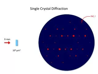
Miller Indices • Miller indices are set of imaginary indices used to identify various points and plans in the unit cell • They are designated as h, k, l and have whole integer values (001, 010, 100, 102, 201, 021, etc) 21
Miller Indices 21 Fundamentals of Powder diffraction and Structural Characterization of Materials, by Vitalij K., et al