Download
1 / 38
400 likes | 802 Views
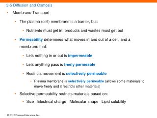
3-5 Diffusion and Osmosis. Membrane Transport The plasma (cell) membrane is a barrier, but: Nutrients must get in; products and wastes must get out Permeability determines what moves in and out of a cell, and a membrane that: Lets nothing in or out is impermeable

E N D
3-5 Diffusion and Osmosis • Membrane Transport • The plasma (cell) membrane is a barrier, but: • Nutrients must get in; products and wastes must get out • Permeability determines what moves in and out of a cell, and a membrane that: • Lets nothing in or out is impermeable • Lets anything pass is freely permeable • Restricts movement is selectively permeable • Plasma membrane is selectively permeable (allows some materials to move freely and it restricts other materials) • Selective permeability restricts materials based on: • Size Electrical charge Molecular shape Lipid solubility
3-5 Diffusion and Osmosis • Membrane Transport • Transport through a plasma membrane can be: • Active(requiring energy and ATP) • Passive (no energy required) • Diffusion (passive) • Carrier-mediated transport (passive or active) • Vesicular transport (active)
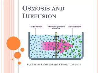
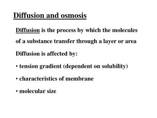
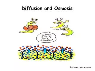
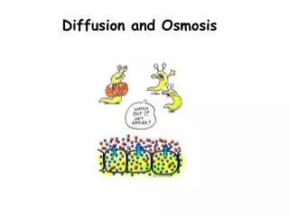
3-5 Diffusion and Osmosis • Diffusion • All molecules are constantly in motion • Molecules in solution move randomly • Random motion causes mixing • Concentration is the amount of solute in a solvent • Concentration gradient • More solute in one part of a solvent than another
3-5 Diffusion and Osmosis • Factors Influencing Diffusion • Distancethe particle has to move • Molecule Size • Smaller is faster • Temperature • More heat, faster motion • Concentration Gradient • The difference between high and low concentrations • Electrical Forces • Opposites attract, like charges repel
3-5 Diffusion and Osmosis Diffusion across Plasma Membranes Can be simple or channel mediated Materials that diffuse through plasma membrane by simple diffusion Lipid-soluble compounds (alcohols, fatty acids, and steroids) • Channel-mediated diffusion • Water-soluble compounds and ions • Factors in channel-mediated diffusion • Size • Charge • Interaction with the channel – leak channels Dissolved gases (oxygen and carbon dioxide)
Figure 3-15 Diffusion across the Plasma Membrane EXTRACELLULAR FLUID Lipid-soluble moleculesdiffuse through theplasma membrane Channelprotein Plasma membrane Small water-solublemolecules and ionsdiffuse throughmembrane channels Large molecules that cannotdiffuse through lipids cannotcross the plasma membraneunless they are transportedby a carrier mechanism CYTOPLASM
3-5 Diffusion and Osmosis • Osmosis: A Special Case of Diffusion • Osmosisis the diffusion of water across the cell membrane • More solute molecules, lower concentration of water molecules • Membrane must be freely permeable to water, selectively permeable to solutes • Water molecules diffuse across membrane toward solution with more solutes • Volume increases on the side with more solutes Identify the effect that a 2% solution of NaCl on RBCs.
3-5 Diffusion and Osmosis • Osmolarity and Tonicity • The osmotic effect of a solute on a cell • Two fluids may have equal osmolarity (solute concentration), but different tonicity (how the solution affects a cell). • Isotonic(iso-= same, tonos= tension) • A solution that does not cause osmotic flow of water in or out of a cell • Hypotonic(hypo- = below) • Has less solutes and loses water through osmosis • Hypertonic(hyper- = above) • Has more solutes and gains water by osmosis
3-5 Diffusion and Osmosis • Osmolarity and Tonicity • A cell in a hypotonic solution: • Gains water • Ruptures (hemolysisof red blood cells) • A cell in a hypertonic solution: • Loses water • Shrinks (crenationof red blood cells)
Figure 3-17 Osmotic Flow across a Plasma Membrane Hemolysis Crenation No change Watermolecules Solutemolecules SEM of normal RBCin an isotonic solution SEM of RBC in ahypotonic solution SEM of crenated RBCsin a hypertonic solution
3-6 Carriers and Vesicles • Carrier-Mediated Transport • Of ions and organic substrates • Characteristics • Specificity • One transport protein, one set of substrates • Saturation Limits • Rate depends on transport proteins, not substrate • Regulation • Cofactors such as hormones • Cotransport (symport) • Two substances move in the same direction at the same time • Countertransport (antiport) • One substance moves in while another moves out
3-6 Carriers and Vesicles • Carrier-Mediated Transport • Facilitated Diffusion (Passive) • Carrier proteins transport molecules too large to fit through channel proteins (glucose, amino acids) • Molecule binds to receptor site on carrier protein • Protein changes shape, molecules pass through • Receptor site is specific to certain molecules
3-6 Carriers and Vesicles • Carrier-Mediated Transport • Active Transport (Primary or Secondary) • Active transport proteins • Move substrates against concentration gradient • Require energy, such as ATP • Ion pumps move ions (Na+, K+, Ca2+, Mg2+) • Exchange pump counter transports two ions at the same time • Primary Active Transport • Sodium–potassium exchange pump • Active transport, carrier mediated Sodium ions (Na+) out (3), potassium ions (K+) in (2) • 1 ATP moves 3 Na+ and 2 K+
Figure 3-19 The Sodium-Potassium Exchange Pump EXTRACELLULARFLUID Sodiumpotassiumexchangepump CYTOPLASM
3-6 Carriers and Vesicles • Vesicular Transport (Bulk Transport) • Materials move into or out of cell in vesicles • Endocytosis (endo- = inside) is active transport using ATP • Receptor mediated • Pinocytosis • Phagocytosis Receptor-mediated endocytosis • Receptors (glycoproteins) bind target molecules (ligands) • Coated vesicle (endosome) carries ligands and receptors into the cell
Figure 3-21 Receptor-Mediated Endocytosis F u s i o n Receptor-Mediated Endocytosis Ligands EXTRACELLULAR FLUID Ligands bindingto receptors Target molecules (ligands) bind toreceptors in plasma membrane. Exocytosis Endocytosis Ligandreceptors Areas coated with ligands formdeep pockets in plasmamembrane surface. Pockets pinch off, formingendosomes known as coatedvesicles. Coatedvesicle Coated vesicles fuse with primarylysosomes to form secondarylysosomes. m e n h t c a t e D Ligands are removed and absorbed into the cytoplasm. Primarylysosome Ligandsremoved Secondarylysosome The lysosomal and endosomalmembranes separate. CYTOPLASM The endosome fuses with theplasma membrane, and thereceptors are again available forligand binding.
3-6 Carriers and Vesicles Endocytosis Pinocytosis (“cell drinking”) Endosomes “drink” extracellular fluid Phagocytosis (“cell eating”) Pseudopodia (pseudo- = false, pod- = foot) Engulf large objects in phagosomes Exocytosis (exo- = outside) Granules or droplets are released from the cell
Table 3-2 Mechanisms Involved in Movement across Plasma Membranes
3-7 Transmembrane Potential • Transmembrane Potential • Charges are separated creating a potential difference • Unequal charge across the plasma membrane is transmembrane potential • Resting potential ranges from –10 mV to –100 mV, depending on cell type
3-8 Cell Life Cycle • Cell Life Cycle • Most of a cell’s life is spent in a nondividing state (interphase) • Body (somatic) cells divide in three stages • DNA replicationduplicates genetic material exactly • Mitosisdivides genetic material equally • Cytokinesis divides cytoplasm and organelles into two daughter cells
3-8 Cell Life Cycle • DNA Replication • Helicases unwind the DNA strands • DNA polymerase • Promotes bonding between the nitrogenous bases of the DNA strand and complementary DNA nucleotides dissolved in the nucleoplasm • Links the nucleotides by covalent bonds • DNA polymerase works in one direction • Ligases piece together sections of DNA
Figure 3-23 DNA Replication DNA polymerase DNA strand unwinds Segment 2 DNA nucleotide KEY Segment 1 Adenine DNApolymerase Guanine Begins attaching complimentary nucleotides Cytosine Thymine
3-8 Cell Life Cycle • Interphase • The nondividing period • G-zero (G0)phase— specialized cell functions only • G1 phase— cell growth, organelle duplication, protein synthesis • S phase— DNA replication and histone synthesis • G2 phase— finishes protein synthesis and centriole replication
Figure 3-24 Stages of a Cell’s Life Cycle: Interphase 6 to 8 hours 2 to 5 hours hours more or 8 s r u ho 3 to 1 SIS NE KI TO CY INTERPHASE Most cells spend only a small part of theirtime actively engaged in cell division.Somatic cells spend the majority of theirfunctional lives in a state known asinterphase. During interphase, a cellperfoms all its normal functions and, ifnecessary, prepares for cell division. When the activities of G1 have been completed, the cell enters the S phase. Over the next 68 hours, the cell duplicates its chromosomes. This involves DNA replication and the synthesis of histones and other proteins in the nucleus. A cell that is ready todivide first enters the G1phase. In this phase, the cell makes enough mitochondria, cytoskeletal elements, endo- plasmic reticula, ribosomes, Golgi membranes, and cytosol for two functionalcells. Centriole replica- tion begins in G1 and commonly continues until G2. In cells dividing at top speed, G1 may last just 812 hours.Such cells pourall their energyinto mitosis, andall other activitiescease. If G1 lastsfor days, weeks, ormonths, preparationfor mitosis occurs as the cells perform their normal functions. Once DNA replication has ended, there is a brief (25-hour) G2 phase devoted to last-minute protein synthesis and to the comple- tion of centriole replication. SDNAreplication,synthesis ofhistones G2Proteinsynthesis G1 Normal cell functionsplus cell growth,duplication of organelles, protein synthesis THECELLCYCLE Centrioles incentrosome Prophase MITOSIS Metaphase Nucleus Anaphase Telophase G0 Interphase MITOSIS ANDCYTOKINESIS An interphase cell in the G0 phase is not preparing for division, but is performing all of the other functions appropriate for that particular cell type. Some mature cells, such as skeletal muscle cells and most neurons, remain in G0 indefinitely and never divide. In contrast, stem cells, which divide repeatedly with very brief interphase periods, never enter G0. Duringinterphase,the DNA strandsare looselycoiled andchromosomescannot be seen.
3-8 Cell Life Cycle • Mitosis • Divides duplicated DNA into two sets of chromosomes. DNA coils tightly into chromatids. Chromatids connect at a centromere. Protein complex around centromere is kinetochore.
3-8 Cell Life Cycle • Mitosis • Prophase • Nucleoli disappear • Centriole pairs move to cell poles • Microtubules (spindle fibers) extend between centriole pairs • Nuclear envelope disappears • Spindle fibers attach to kinetochore • Metaphase • Chromosomes align in a central plane (metaphase plate)
Figure 3-24 Stages of a Cell’s Life Cycle: Mitosis and Cytokinesis Chromosomewith two sisterchromatids Chromosomalmicrotubules Metaphaseplate Astral rays andspindle fibers Centrioles(two pairs) Late prophase Metaphase Early prophase
3-8 Cell Life Cycle • Mitosis • Anaphase • Microtubules pull chromosomes apart • Daughter chromosomes group near centrioles • Telophase • Nuclear membranes re-form • Chromosomes uncoil • Nucleoli reappear • Cell has two complete nuclei
Figure 3-24 Stages of a Cell’s Life Cycle: Mitosis and Cytokinesis Daughterchromosomes Cleavagefurrow Daughtercells Telophase Cytokinesis Anaphase
3-8 Cell Life Cycle Cytokinesis (Division of Cytoplasm) Cleavage furrow around metaphase plate Membrane closes, producing daughter cells.
Biol 2401 Watch this video on Mitosis: http://www.youtube.com/watch?v=NR0mdDJMHIQ&feature=related
3-8 Cell Life Cycle • The Mitotic Rate and Energy Use • Rate of cell division • Slower mitotic rate means longer cell life • Cell division requires energy (ATP) • Muscle cells, neurons rarely divide • Exposed cells (skin and digestive tract) live only days or hours – replenished by stem cells
3-10 Cell Division and Cancer Cancer Developes in Steps Abnormal cells → Primary tumor → Mestasis → Secondary tumor • Tumor(Neoplasm) • Enlarged mass of cells • Abnormal cell growth and division • Benign tumor • Contained, not life threatening unless large • Malignant tumor • Spreads into surrounding tissues (invasion) • Starts new tumors (metastasis)
Figure 3-25 The Development of Cancer Abnormalcell Primary tumor cells Secondary tumor cells Growth of bloodvessels into tumor Celldivisions Celldivisions Invasion Penetration Escape Circulation
3-11 Differentiation Differentiation All cells carry complete DNA instructions for all body functions Cells specialize or differentiate To form tissues (liver cells, fat cells, and neurons) By turning off all genes not needed by that cell All body cells, except sex cells, contain the same 46 chromosomes Differentiation depends on which genes are active and which are inactive
Biol 2401 TRANSCRIPTION • If the bases of one side of DNA read: A-G-C-T, the complementary (opposite) DNA strand reads: • A-C-G-T. • A-G-A-T. • U-C-G-A. • T-C-G-A. d. T-C-G-A • The bases in a strand of DNA READ: T-C-C-A. The transcribed strand • Of mRNA reads: • A-G-G-T. • G-C-G-A. • U-C-G-T. • A-G-G-U. d. A-G-G-U
Genetic Code GUCCCGUG AUG CCG AGU UGG AGU AGA UAA CUCAGAAU STARTmethionine proline serine tryptophane serine arginine STOP