Download
1 / 88
890 likes | 1.06k Views
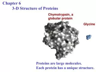
Chapter 6 Proteins in Action. 1. Hemoglobin is a multisubunit allosteric protein that carries O2 in erythrocyte. 1.1 Hemoglobin is a well-studied and well-understood protein. 1.1.1 It was one of the first proteins to have its molecular mass accurately determined.

E N D
1. Hemoglobin is a multisubunit allosteric protein that carries O2 in erythrocyte. 1.1 Hemoglobin is a well-studied and well-understood protein. 1.1.1 It was one of the first proteins to have its molecular mass accurately determined. 1.1.2 The first protein to be characterized by ultracentrifuge. 1.1.3 The first protein to be associated with a specific physiological function.
1.1.4 The first protein with a single amino acid substitution being related to a genetic disease (the beginning of molecular medicine). 1.1.5 The first multisubunit protein with its detailed atomic structure determined by X-ray crystallography. 1.1.6 The best understood allosteric protein.
1.2 Determination of the atomic structure of hemoglobin A (from normal adult) is very revealing. 1.2.1 The protein molecule exists as a a2b2 tetramer. 1.2.2 Each subunit has a structure strikingly and unexpectedly similar to each other and to that of myoglobin, indicating quite different amino acid sequences can specify very similar 3-D structures. 1.2.3 Extensive interactions exist between the unlike subunits through noncovalent interactions. 1.2.4 Quaternary structure changes markedly when O2 binds. Crystals of deoxyhemoglobin shatter (break) when exposed to O2. 1.2.5 O2 binds to the sixth coordination position of the ferrous iron (as in myoglobin).
1.3 Hemoglobin is a much more intricate and sentient (sensitive) molecule than is myoglobin. 1.3.1 The oxygen-binding (dissociation) curve of hemoglobin is sigmoidal and that of myoglobin is hyperbolic. 1.3.2 Myoglobin has a higher affinity for O2, evolved for O2 storage. 1.3.3 Hemoglobin releases O2 efficiently at low oxygen level tissues (evolved for O2 delivery), myoglobin does not. 1.3.4 Oxygen binding of hemoglobin shows positive cooperativity. The binding of O2 (the ligand) at one heme facilitates the binding of O2 at the other hemes on the same tetramer (vice versa, unloading of oxygen at one heme facilitates the unloading of oxygen at the others). (Negative cooperativity refers to a decrease of activity.)
1.3.5 Increasing concentrations of H+ (with a decrease of pH) or CO2 lowers the O2 affinity of hemoglobin (H+ and CO2 has no effect on O2 affinity of myoglobin). This is called Bohr effect, which helps the release of O2 in the capillaries of actively metabolizing tissues. (melecular mechanism?) 1.3.6 One molecule of 2,3-diphosphoglycerate (BPG) binds to the central cavity of one tetramer of hemoglobin, which lowers its O2 affinity. 1.3.7 Fetal hemoglobin (HbF) binds BPG less strongly than does hemoglobin A (adult) and consequently has a higher oxygen affinity. (physiological function? Extraction of O2 from the mother)
1.4 Cooperativity is a particular case of an allosteric effect 1.4.1 Allosteric effect refers to the phenomenon in which a molecule (allosteric effector) bound to one site on a protein causes a conformational change in the protein such that the activity of another site on the protein is altered (increased or decreased). 1.4.2 H+, CO2, and BPG all show an allosteric effect (heterotrophic?) for the O2 binding process of hemoglobin.
1.5 Two models have been proposed to explain the allosteric regulation phenomena 1.5.1 The sequential model (proposed by Daniel Koshland, Jr.) hypothesizes that the binding of one ligand to one subunit changes the conformation of that particular subunit from the T state (with a low activity) to the R state (with a high acitvity), a transition that increases the activity of the other subunits for the ligand. 1.5.2 The sequential model can be analogized to the tearing process of postage stamps. (see fig.)
1.5.3 The concerted model (proposed by Monod, Wyman, and Changeux) hypothesizes that symmetry is conserved in allosteric transitions (all subunits are in the same conformation) and the binding of each ligand increases the probability that all subunits in that molecule are converted to the R-state (with a high activity). All-or-none model. 1.5.4 The interplay between these different ligand-binding sites is mediated primarily by changes in quaternary structure. The contact region between two subunits can serve as a switch that transmits conformational changes from one subunit to another.
T: tense state, circle, less active; R: relaxed state, square, more active Concerted, all-or-none Sequential
1.5.5 The functional characteristics of an allosteric protein are regulated by specific molecules in its environment. In another words, in the evolutionary transition from myoglobin to hemglobin, a macromolecule capable of perceiving information from its environment has emerged.
1.6 Sickle-cell anemia was found to be caused by a single amino acid change in the b chain of hemoglobin molecules. 1.6.1 The hemoglobin molecule from sickle-cell anemia patients (HbS) was found to have a higher pI value (having more net positive charges). 1.6.2 Peptide fingerprinting (protease digestion + electrophoresis + chromatography) of HbS and HbA (wt) revealed that all but one of the peptide spots matched. 1.6.3 Amino acid sequencing revealed that HbS contains Val instead of Glu is at position 6 of the b chain! 1.6.4 The oxygen binding affinity and allosteric properties of hemoglobin are virtually unaffected by this change (the b6 is located at the surface of the protein).
1.6.5 High concentration of deoxygenated HbS forms fiber participatates, which sickles the red blood cells, because the fiber formation is a highly concerted reaction. 1.6.6 Presence of Val6 on the b subunits generates a hydrophobic patch on the surface which complements with another hydrophobic patch formed only in deoxygenated HbS, thus generating the fiber precipitates (a polymer of HbS). 1.6.7 Sickle cell trait (heterozygote) confers a small but highly significant degree of protection against the most lethal form of malaria (probably by accelerating the destruction of infected erythrocytes, in Africa). 1.6.8 Fetal DNA can be analyzed for the presence of the HbS gene (prenatal DNA diagnosis).
1.7 Thalassemias are genetic disorders characterized by defective synthesis of one or more hemoglobin chains. 1.7.1 This can be caused by a missing gene, impaired RNA synthesis or processing, generation of grossly abnormal proteins.
binding sites occupied [L] = ------------------------------- = ------------- total binding sites [L] + Kd is a measurable quantity in experiment. [L] is the free ligand concentration.
Bohr’s effect and its molecular mechanism Release of O2 in peripheral tissues Binding of O2 in lungs with release of H+.
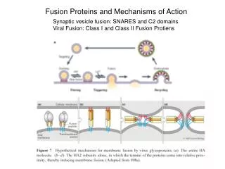
2. Immunoglobulin superfamily members, found on cell surfaces or secreted, are widely used for specific molecular recognition. 2.1 An immunoglobulin G (IgG) molecule (of ~150 kD) was found to contain two light and two heavy chains, connecting to each other through disulfide bonds. 2.1.1 Papain digestion convert the IgG molecule into two Fab and one Fc fragments. 2.1.2 Each Fab fragment (containing one complete light chain and half of a single heavy chain) binds one molecule of antigen in a similar manner to the original immunoglobulin molecule.
2.1.3 Each IgG molecule binds to two molecules of antigens (thus called bivalent). 2.2 Complete amino acid sequence analysis of purified myeloma patient’s immunoglobulins revealed strikingly that the L and H chains consist of variable and constant regions. 2.2.1 Residues 1 to 108 in the L chains are relatively variable, and 109 to 204 relatively constant. 2.2.2 Residues 1 to 108 in the H chains are variable, and 109 to 446 relatively constant. 2.2.3 Three segments in the L chain and three in the H chain display far more variability than do others, which are thus named as hypervariable segments (also called complementary-determining regions, or CDRs, because they determine antibody specificity).