Download
1 / 8
90 likes | 552 Views
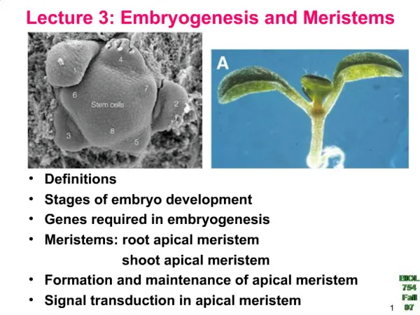
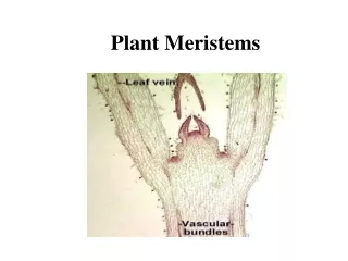
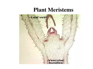
Root meristems and primary tissues. Root apical meristem mitosis. Be able to identify all of the mitotic stages and know what order they occur in. (review). Root promeristems. apical meristem promeristems primary tissues. Root promeristems. What tissues are forming here?.

E N D
Root apical meristem mitosis Be able to identify all of the mitotic stages and know what order they occur in. (review)
Root promeristems apical meristem promeristems primary tissues
Root promeristems What tissues are forming here?
Dicot root primary tissuesYou should be able to identify the tissues and cell types and know the functions of the cell types. Be able to identify epidermis, cortex, parenchyma, endodermis, passage cells, stele or vascular cylinder, pericycle, xylem, phloem,residual procambium, casparian strips.
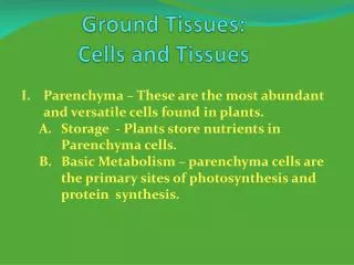
Starch grains in parenchyma cells Parenchyma cells are where plants store most of their starch. When proplastids in parenchyma cells are exposed to light, they often develop into chloroplasts. Plants usually store more starch in their roots than in their stems. Can you think of a possible reason why they do that?
Monocot root primary tissues Note that there is parenchyma in the middle of this monocot root. Cells in the outer part of the cortex have differentiated into fibers, and the epidermis is disappearing. Be able to identify epidermis, exodermis, cortex and cortical parenchyma, endodermis and endodermal cells, pericycle, primary phloem, primary xylem, vessel cells, sieve cells, undifferentiated xylem parenchyma. (This is a cross section of a corn or maize root, Zea.)
Root anatomy of another monocot root Notice that the endodermis is very lignified. Do you think water could pass through this endodermis?) Be able to identify all tissues and cell types. (These are Smilax root cross sections. The common Name of Smilax is catbrier or sawbrier.)