Download
1 / 28
290 likes | 335 Views
This comprehensive guide covers DNA cloning, PCR, RFLP, restriction enzymes, and more in molecular genetics diagnosis methods. Learn about in-vivo cell-based mechanisms, recombinant DNA technology, and the importance of restriction mapping in DNA manipulation techniques.

E N D
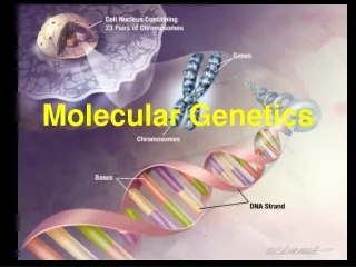
Molecular Genetics Diagnosis Methods By: Mahdi Bijanzadeh MD, PhD. bijanzadeh-m@ajums.ac.ir
Molecular Genetics Diagnosis Methods At the end of this session, you should answer this questions: 1. How many types of DNA cloning are there? 2. What does PCR carried out? 3. How does PCR product analysis? 4. What does RFLP carried out?
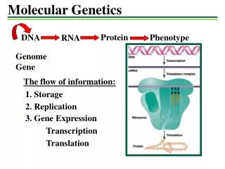
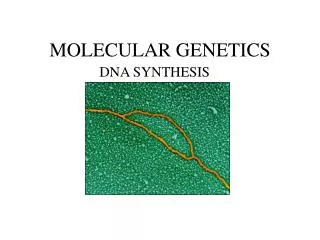
DNA cloning or DNA synthesis The selective amplification of a specific DNA fragment or sequence to produce relatively large amounts of ahomogeneous DNA fragment. Types: 1. Natural, in-vivo cell-based mechanism. 2. Cell-free, in-vitro PCR
In-vivo cell-based cloning Recombinant DNA technology: use in DNA cloning. The key to cloning a DNA fragment of interest is to link it to a vector DNA molecule that can replicate within a host cell.
In-vivo cell-based cloning Six basic steps: 1. Generation of DNA fragments: enzymes of certain microbes can cleave double stranded DNA in or near a particular sequence of nucleotides: restriction enzymes or endonuclease, 300: a staggered (sticky or cohesive) or a blunt end. Restriction sites: specific 4-8 bp sequence recognized by .restriction enzymes →restriction fragments
Restriction enzymes *Phage (or viruses) invade all types of cells. Bacteria are one favorite target. *Defense mechanisms have been developed by bacteria to defendthemselves from these invasions. Bacteria have evolved a class of enzymes that destroy foreign DNA (eg. virus DNA): protect bacteria from bacteriophages. *Infecting DNA is cleaved (restricted) bythe restriction enzyme(s) preventing it from successfully replicating and parasitizing the cell.
Why the bacteria does not kill itself? *Usually, organisms that make restriction enzymesalso make a companion modification enzyme(DNA methyltransferase-methylase) that protects their own DNA from cleavage. *These enzymes recognize the same DNAsequence as the restriction enzyme accompany, but instead of cleaving the sequence,they disguise it by methylating one of the bases in each DNA strand. *Restriction enzyme system is composed of a restriction endonuclease enzyme and a methylase enzyme. *Each bacterial species and strain has their own combination of restriction and methylating enzymes.
Enzyme activity Scanning GGACGCTAGCTGATGAATTCGCATCGGATCCGAATCCGCTCTTTCAA CCTGCGATCGACTACTTAAGCGTAGCCTAGGCTTAGGCGAGAAAGTT Recognition Sequence GGACGCTAGCTGATGAATTCGCATCGGATCCGAATCCGCTCTTTCAA CCTGCGATCGACTACTTAAGCGTAGCCTAGGCTTAGGCGAGAAAGTT Cleavage AATTCGCATCGGATCCGAATCCGCTCTTTCAA GCGTAGCCTAGGCTTAGGCGAGAAAGTT GGACGCTAGCTGATG CCTGCGATCGACTACTTAA
Nomenclature Smith and Nathans (1973) proposed enzyme naming scheme; * Three-letter acronym for each enzyme derived from the source organism * First letter from genus * Next two letters represent species * Additional letter or number represent the strain or serotypes. For example: the enzyme HindII was isolated from Haemophilus influenzae serotype d.
Ends of restriction fragments: 1. Blunt ends 2. Sticky ends: Most RE.s make staggered cuts. • 3‘extensions • 5‘extensions 5' termini of each strand in thecleavage product(s) retain the phosphoryl group from the phosphodiester bond, the 3' termini are hydroxylated.
Star effect: Optimum conditions are necessary for the expected result. Under extreme conditions such as elevated pH or low ionic strength, restriction enzymes are capable of cleaving sequences which are similar but not identical to their recognition sequence. EcoR1→GAATTC EcoR1 with star activity→NAATTN (N=any base)
Restriction mapping *A method to map an unknown segment of DNA by breaking it by restriction enzymes, into pieces and then identifying the locations of the breakpoints. After a DNA segment has been digested, the resulting fragments can be separated using gel electrophoresis. *The DNA to be restriction mapped it usually contained within a well-characterized plasmid or bacteriophage vector for which the sequence is known. In fact, there are usually multiple known restriction sites immediately flanking the uncharacterized DNA, which facilitates making the map.
An example You have isolated a clone in pBluescript. You know how big it portion of the plasmid is (3.0 kb) and what RE.s are present in the plasmid (from the company). You also know that the insert is 2.0 kb long & that it is inserted the Eco RI site. Your task is to find out more information about the insert:
You would digest plasmid with an enzyme that you know is in the pBluescript plasmid. For example, you know 1 Bam HI site is there, in the multiple cloning site next to the Eco RI site. Digestion with Bam HI: 2 possibilities: 1) no Bam HI sites in the insert: you see only one DNA fragment~5.0 kb long (3.0 kb of pBluescript DNA & 2.0 kb of insert DNA). 2) A Bam HI site in the insert: enzyme will cut the circular plasmid in 2 places: 2 fragments of DNA, their sizes (determined by electrophoresis) will tell you where the site is.
Uses of Restriction Mapping It is important for many techniques for manipulate DNA: - To cut large piece of DNA into smaller fragments to allow it to be sequenced. Genes & cDNAs can be 1000 kb or Mb long; but they can only be sequenced 400 bases at a time: they must be chopped up into smaller pieces and subcloned to perform the sequencing. - An easy way to compare DNA fragments without having any information of their nucleotide sequence. For example, you may isolate 2 clones for a gene that are 8 kb & 10 kb long. You know that they overlap, because the procedure you used to isolate them told you that they have sequences in common. A restriction map can tell you how much they overlap by:
2. Recombination of DNA fragments: Sticky or Cohesive ends will unite under appropriate conditions with complementary sequences produced by the same RE on DNA from any source. DNA ligase: covalently joins the ends of the restriction fragment and vector DNA by 3’ →5’ phosphodiester bonds (after first hydrogen bonding), then inserted into vector DNA.
3. Vectors or replicon; with incorporation of the foreign or target DNA into a vector allows the production of large amounts of that DNA fragment. Modification to ensure the insertion: generally resistance to AB. Competent: bacterial cell membrane is not permeable to large molecules such as DNA fragments, but can be made permeable by exposure to certain salts or high voltage. Five types: plasmids, bacteriophages, cosmid, bacterial and yeast artificial chromosomes (BAC & YAC).
4. Transformation of the host organism: produce the large quantities of identical copies of the original single target DNA or clones.
5. Screening for recombinant vectors: by a detection system; for example, loss of antibiotic resistance. 6. Selection of specific clones: nucleic acid hybridization (blot studies) or with an available antibody.
DNA Libraries Sources of DNA used to make recombinant DNA molecules: 1. DNA from nucleated cells: total or genomic DNA. 2.DNA made by the action of the reverse transcriptase on mRNA: complementary DNA or cDNA. By using a specific tissue or cell type as a source of mRNA: immature RBC(reticulocytes) containing predominantly globin mRNA resulted in cloning of the genes for the globin chains of hemoglobin. DNA library: The collection of recombinant DNA molecules generated from a specific source, for example a genomic or cDNA library.
Cell free DNA cloning Polymerase Chain Reaction (PCR) - For generating unlimited amounts of a sequence of interest. - It can selectively amplify a single molecule of DNA several billion-fold in a few hours. - Definition: An enzymatic amplification of a fragment of DNA (the target) located between two oligonucleotide “primers”. - A fast, cheep & easy method
PCR - The newly synthesized strand of DNA are themselves complementary and can form a second copy of the original target sequence → exponential amplification (2, 4, 8, 16, 32, …copies) until the substrates are used up. - Amplified segments can be sequenced/ tested by ASO hybridization to detect a mutation/ evaluated.
How many types of DNA cloning are there? What does PCR carried out? How does PCR product analysis? What does RFLP carried out?