Download
1 / 1
10 likes | 200 Views
18F)FDG-PET/CT METABOLIC CHARACTERISATION OF LUNG NODULES IN PAEDIATRIC PATIENTS A.Cistaro 1 , L. Gastaldo 2 , M. Berta 3 , A. Brach del Prever 3 , G. Di Rosa 4 , C. De Filippi 4 , M. Mancini 1. 1. Positron Emission Tomography IRMET S.p.A., Turin, Italy

E N D
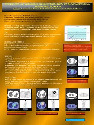
18F)FDG-PET/CT METABOLIC CHARACTERISATION OF LUNG NODULES IN PAEDIATRIC PATIENTS A.Cistaro1, L. Gastaldo2, M. Berta3, A. Brach del Prever3, G. Di Rosa4, C. De Filippi4, M. Mancini1 1. Positron Emission Tomography IRMET S.p.A., Turin, Italy 2. Department of Paediatric Sciences, Regina Margherita Infant Hospital, Turin, Italy 3. Onco-Hematology Department, Regina Margherita Infant Hospital, Turin, Italy 4.Operative Unit of Radiology, Regina Margherita Infant Hospital, Turin, Italy PET/CT is successfully used in metabolic characterisation of solitary lung nodule in adult patients. An SUV max of 2.5 is generally accepted to distinguish benign from malignant nodules. Very few studies have been performed in paediatric patients. AIM Individuating the lowest dimension (visual analysis) and the lowest semi-quantitative value capable to metabolically characterise lung nodules in paediatric patients MATERIAL AND METHODS 63 patients were enrolled: 32 patients: 12 osteosarcomas, 13 Ewing’s sarcomas, 2 HL, 1 NHL, 1 EBV-related Lymphohistiocytosis, 1 pinealoblastoma, 1 ALL. 84 lung nodules have been studied. PET/CT results have been compared to histology and clinical-radiological follow-up. Distribution of SUVmax for all nodules. In blue colour are represented metastasis and in pink the benign lesions. It seems to be an SUVmax value able to differentiate metastases from benign lesions. The value is around 1. RESULTS Visual analysis: sensitivity 90,1%; specificity 97,5%; PPV 97,5%; NPV 90,7%; accuracy 94%(p<0,05). Semi-quantitative analysis with an SUVmax cut-off >= 1: sensitivity 93,3%; specificity 87,1%; PPV 89,3%; NPV 91,8%; accuracy 90,4%. Restricting the same analysis (SUVmax >= 1) to nodules with a diameter higher than 5 mm the sensitivity was >99%; specificity 69,2%; accuracy 92,7%. No statistically significant results obtained for nodules with diameter < 5mm. The same analysis performed with an SUVratio>=1 (SUVratio = SUVmax lesion/SUVmax mediastinic (measured on aortic arch) obtained similar results. CONCLUSIONS PET/CT seems accurate and sensitive for the metabolic characterisation of lung nodules higher than 5 mm of diameter also in paediatric patients. An SUVmax and an SUVratio >= 1 seem an useful semi-quantitative cut-off, capable to improve the metabolic characterisation of lung nodules. Micotic lung lesion in pazient affected by lymphoistiocytosis EBV correlated Lung metastasis from osteosarcoma Lung metastasis from adrenal gland carcinoma Lung localitation of LNH For contact: a.cistaro@irmet.com