Download
1 / 87
880 likes | 1.1k Views
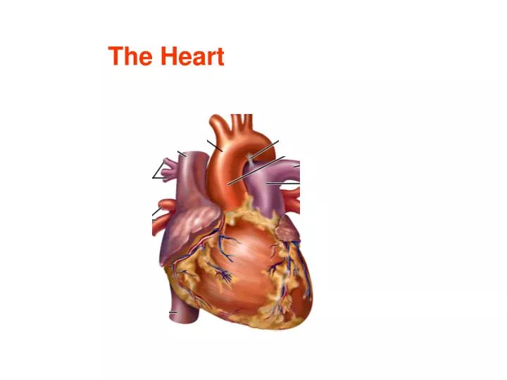
The Heart. Chambers of the Heart. Cardiac Cycle Ventricular systole - isovolumic contraction - ejection Ventricular diastole - isovolumic relaxation - rapid filling - atrial contraction. Isovolumic Ventricular Contraction. 2) Ventricular Ejection.

E N D
Cardiac Cycle Ventricular systole - isovolumic contraction - ejection Ventricular diastole - isovolumic relaxation - rapid filling - atrial contraction
Isovolumic • Ventricular Contraction 2) Ventricular Ejection 3) Isovolumic Ventricular Relaxation 4) Ventricular Filling 5) Atrial Contraction
Autorhythm The heart can beat on its own without the need for exogenous commands.
Conclusion ? The heart generates electricity. Motor nerve Skeletal muscle
TERMINOLOGY • Excitation • - definition: generation of action potentials • - different from contraction • Contraction • - definition: shortening of muscle cells • - triggered by excitation
Excitation-Contraction coupling [ Ca++ ]i Contraction Excitation (Action Potentials) (shortening)
Sequence of excitation Sinus-Atrial node (SA node) Atria Atrial-ventricular node (AV node) Ventricles
Original Impulses from S-A Node The electrical impulses are normally generated by a group of specialized pacemaker cells at sinoatrial (SA) node. SA node - located in the right atrial wall, just inferior to the entrance of the superior vena cava.
Conduction of Action Potentials from Cell to Cell • through gap junctions in intercalated discs • (electrical synapses)
Conduction in Atria The electrical impulses from SA node spread through the entire right and left atrial muscle mass, triggering contraction of the right and left atrium.
Delay at A-V Node - The impulses from S-A node travel to atrioventricular (A-V) node. - A-V node is located in lower end of the interatrial septum near the tricuspid valve. A-V node
Delay at A-V Node - Conduction speed in A-V node is slow (delay). - This delay allows time for the atria to finish contraction and empty their contents into the ventricles before ventricles start to contract. - A-V node is the only normal route that impulses from SA node are transmitted into ventricles.
From AV node to Ventricles • His bundle • left branch (anterior/posterior division) • - right branch His bundle
Rapid Conduction in Ventricles After the delay at A-V node, the impulses rapidly spread to the ventricles via specialized fibers, Purkinje fibers. • 1) Purkinje fibers • located in the subendocardial layer • - fastest conduction (4 m/s) • 2) Ordinary ventricular myocardial cells • able to conduct AP at a slower speed
Rapid conduction in the ventricles simultaneous excitation of the ventricles functional syncytium
NNote: - Each electrical impulse can trigger cardiac muscle contraction normally only once. - A normal heart generates 60 to 100 impulses in 1 minute at resting state. 1 1
Properties of Cardiac Muscle Excitation of the heart is triggered by electrical impulse rather than neural transmitters. Contraction of the heart is triggered by elevation of intracellular calcium influx. [ Ca++ ]i Contraction Excitation (Action Potentials) (shortening)
Properties of Cardiac Muscle - Myocytes depend heavily on oxygen and blood supply. - Not fatigue - Excitability Cycle The myocytes have Long refractory periodduring which they do not respond to any electrical impulses.
RRole of a Long Refractory Period – 1 prevent ventricles from contracting at too high rates so that enough time is allowed for refill of the ventricles
Role of Long refractory period - 2 Prevent retrograde excitation
ELECTROCARDIOGRAPHY (ECG)
EELECTROCARDIOGRAPHY ((ECG) the recording of electrical activities of the heart via electrodes placed on body surface.
Applications of ECG • 1) measure automaticity • HR, rhythmicity, pacemaker • 2) measure conductivity • pathway, reentry, block • 3) reveal hypertrophy • 4) reveal ischemic damages • location, size, and progress
Waves and Intervals of ECG P wave: atrial depolarization QRS complex: ventricular depolarization T wave: ventricular repolarization
Disorders of the Cardiac Conduction System ---- Arrhythmias - refers to abnormal initiation or conduction of electrical impulses in the heart. - caused by ischemia, fibrosis, inflammation, or drugs.
Bradycardia • slow heart rate ( < 60 beats/min) • Tachycardia • fast heart rate ( > 100 beats/min)
Atrial or VentricularFlutter and Fibrillation - contract uncoordinatedly and extremely rapidly. - Ventricular fibrillation is lethal.
Premature contraction is when the heart beat is triggered by ectopic pacemakers (cells other than SA node).
Artificial Pacemaker Application: sinus abnormality, complete AV or ventricular block Function: - generate electric pulses - sensing - antitachyarrhythmia
Heart Sounds Four heart sounds can be recorded via phonocardiography, but normally only two, the first and the second heart sounds, are audible through a stethoscope.
First heart sound: • occurs when the atrioventricular (AV) valves closeat the beginning of ventricular contraction. • generated by the vibration of the blood and the ventricular wall • is louder, longer, more resonant than the second heart sound.
Second heart sound - occurs when aortic and pulmonary semilunar valves close at the beginning of ventricular dilation - generated by the vibration of the blood and the aorta - Aortic valve closes slightly before pulmonary valve.
Heart Murmur - abnormal heart sound - occur in valvular diseases and septal defects
Two Basic Types of Valvular Diseases 1) valvular stenosis, a narrowing of the valve 2) valvular insufficiency (incompetence). A valve is unable to close fully; so there is some backflow (regurgitation) of blood.
MECHANICAL PROPERTIES OF THE HEART • CONTENT • Heart Rate • Stroke volume • Cardiac Output (CO) • Ejection Fraction • Preload • Afterload • Contractility • Frank-Starling Mechanism • Factors on Cardiac Output
Heart Rate the number of heart beats in 1 minute. Normal value: 60-100/min Stroke volume the volume of blood pumped out by each ventricle per each contraction. SV
70 ml 75 beat/min Cardiac Output (CO) the amount of blood pumped out by each ventricle in 1 minute. Cardiac output = stroke volume x heart rate Example: 70 ml x 75 beat/min = 5,250 ml/min
SV = 70 ml 130 ml End of diastole End of systole Ejection Fraction = stroke volume end-diastolic ventricular volume 70 ml 130 ml = 54% 60 ml
increases during exercise 120 ml 133 ml = 90% Ejection Fraction SV = 120 ml 133 ml End of diastole End of systole
Preload • the force that stretches the muscle before contraction. • Afterload • the force that stretches muscle during contraction. afterload preload
Preload to ventricles = ventricular end diastolic pressure - the degree of stretch of the ventricular muscle cells just before they contract. - determined by ventricular filling.
Afterload to left ventricle: aortic arterial pressure Afterload to right ventricle: pulmonary arterial pressure Afterload to the left ventricle is greater than that to the right ventricle. Aortic arterial pressure
Contractility - the intrinsic strength of cardiac muscles.
Factors on Cardiac Output • Preload: • 2) Afterload: • 3) Contractility: • 4) Heart Rate: