Download

1 / 47
630 likes | 2.2k Views
Proteins and Electrophoresis. Roger L. Bertholf, Ph.D. Associate Professor of Pathology Director of Clinical Chemistry & Toxicology. Protein Trivia. The most abundant organic molecule in cells (50% by weight) 30-50K structural genes code for proteins

E N D
Proteins and Electrophoresis Roger L. Bertholf, Ph.D. Associate Professor of Pathology Director of Clinical Chemistry & Toxicology
Protein Trivia • The most abundant organic molecule in cells (50% by weight) • 30-50K structural genes code for proteins • Each cell contains 3-5K distinct proteins • About 300 proteins have been identified in plasma
Functional diversity of proteins • Structural • Keratin, collagen, actin, myosin • Transport • Hemoglobin, transferrin, ceruloplasmin • Hormonal • Insulin, TSH, ACTH, PTH, GH • Regulatory • Enzymes • What else?
The composition of proteins • Amino acids (simple proteins) • 20 common (standard) amino acids • Conjugated proteins contain a prosthetic group: • Metalloproteins • Glycoproteins • Phosphoproteins • Lipoproteins • Nucleoproteins
The size of proteins • An arbitrary lower limit is a MW of 5,000 • Proteins can have MW greater than 1 million, although most proteins fall in the range of 12-36K • 100-300 amino acids • Albumin (the most abundant protein in humans) is 66K and contains 550 amino acids (residues)
Protein structure • Primary structure • Amino acid sequence • Secondary structure • -helix or random coil • Tertiary structure • 3-D conformation (globular, fibrous) • Quaternary structure • Multi-protein assemblies
Amino acids (1º structure) • The amino acid sequence is the only genetically-stored information about a protein • Each amino acid is specified by a combination of 3 nucleic acids (codon) in mRNA: • e.g., CGU=Arg; GGA=Gly; UUU=Phe
Undissociated form Zwitterion (dipolar) Properties of amino acids • The –R group determines, for the most part, the properties of the amino acid • Substances that can either donate or accept a proton are called ampholytes
pK2 pK1 Acid-base properties of amino acids pH pKI equivalents OH-
Acidic and basic amino acids • Acidic • Asp R=CH2COO- • Glu R=(CH2)2COO- • Basic • Lys R=(CH2)4NH3+ • Arg R= (CH2)3NHC(NH2)2+ • His R:
Uncharged amino acids • Non-polar (hydrophobic) amino acids • Ala, Val, Leu, Ile, Pro, Phe, Trp, Met • Polar (hydrophilic) amino acids • Gly, Ser, Thr, Cys, Tyr, Asn, Gln
Stereochemistry of amino acids • All naturally-occurring amino acids found in proteins have the “L” configuration L-Alanine D-Alanine
Essential amino acids • Humans ordinarily cannot synthesize: • Leu, Ile, Val, Met, Phe, Trp, Thr, Lys, His (Arg) • Dietary protein is the principal source of essential amino acids
The peptide bond H2O
The peptide bond Dipeptide
Amino acid composition and protein properties • The –R groups determine, for the most part, the properties of the protein • Proteins rich in Asp, Glu are acidic (albumin is an example) • Post-translational modifications of proteins have significant effects on their properties, as well.
Coiling (2 structure) • Linus Pauling described the -helical structure of proteins • Pro and OH-Pro break the -helix • Ser, Ile, Thr, Glu, Asp, Lys, Arg, and Gly destabilize the -helix
Folding (3 structure) • J. C. Kendrew deduced the structure of myoglobin from X-ray crystallographic data • Globular proteins have stable 3-dimensional conformations at physiological pH, temperature (Why?)
Myoglobin • Protein 3 structure is influenced by and regions • Proteins fold in order to expose hydrophilic regions, and sequester hydrophobic regions
4 structure • Hemoglobin has 4 subunits • Two chains • Two chains • Many enzymes have quaternary structures
Measuring proteins • By reactivity • Biuret reaction, Lowry method • By chemical properties • Absorption at =260 nm (Phe) or 280 nm (Tyr, Trp) • By activity • Enzymes, immunoglobulins • By immunogenicity
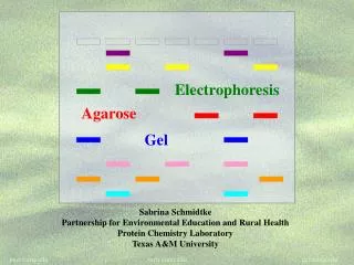
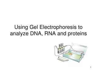
Separating plasma proteins • Chromatography • Gel (size exclusion), HPLC, ion exchange, immunoaffinity • Electrophoresis • Starch gel, agarose gel, cellulose acetate, PAGE
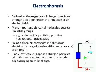
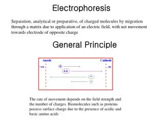
+ – Electrophoresis: Theoretical aspects Electromotive force (emf) + Drag
- - - - - - - - - - - - + – Endosmosis • Large, highly charged proteins may actually migrate toward the like-charged electrode +
Optimizing electrophoresis • Optimal electrophoretic separations must balance speed and resolution • Higher voltage increases speed, but heat causes evaporation of the buffer and may denature proteins • Higher ionic strength (buffer) increases conductivity, but enhances endosmotic effects
Albumin 1 2 + - Serum protein electrophoresis
Albumin • Most abundant protein in plasma (approximately half of total protein) • Synthesized in liver • t½=15-19 days • Principal functions • Maintaining fluid balance • Carrier • Anti-oxidant activity • Buffer
Clinical significance of albumin • Hyperalbuminemia is rare and of no clinical significance • Hypoalbuminemia • Increased loss (nephrotic syndrome) • Decreased production (nutritional deficit, liver failure) • Analbuminemia • Bisalbuminemia, dimeric albumin
Pre-albumin • Thyroxine-binding protein (not an incipient form of albumin), also called transthyretin, or TBPA • Also complexes with retinol-binding protein (RBP) • Only protein that migrates anodal to albumin • Sensitive marker of nutritional status, since its t½ is only 2 days
1-Antitrypsin • Protease inhibitor that binds to, and inactivates, trypsin • Deficiency is associated with • Pulmonary emphysema • Cirrhosis • SPE is only a screening test for AAT deficiency
Other 1 proteins • 1-Acid glycoprotein (orosomucoid) • Biological function is unknown • 1-Fetoprotein (AFP) • Principal fetal protein, used to screen for fetal abnormalities (neural tube defects)
2-Macroglobulin • Largest non-immunoglobulin in plasma • Protease inhibitor • Increased in nephrotic syndrome (size) • Complete genetic deficiency is unknown
(2) Ceruloplasmin • Copper transport protein • Participates in plasma redox reactions • Cp levels fluctuate with a variety of physiological states, but measurement is usually to screen for Wilson’s disease • Plasma Cp is decreased due to inhibition of synthesis
(2) Haptoglobin • Binds to, and preserves, hemoglobin but not myoglobin • Complex also has peroxidase activity, and may be involved in inflammatory response • Hemolytic diseases can deplete Hp levels
() Transferrin • Iron transport protein, and also binds copper • Transferrin is increased in iron deficiency anemia, as well as pregnancy and estrogen therapy • Decreased in inflammation, malignancy, or liver disease
2-Microglobulin • Small protein (MW=11.8K) • BMG is filtered in the glomerulus, but is reabsorbed in the renal tubules. • Urinary BMG levels are a sensitive measure of renal tubular function • Increased in renal failure
() Compliment proteins • C3 and C4 migrate in the region • Compliment proteins are decreased in genetic deficiencies, and increased in inflammation.
Region • Includes immunoglobulins (IgG, IgA, IgM) and C-reactive protein • Single sharp peak is indicates a paraprotein associated with a monoclonal gammopathy (multiple myeloma) • CRP is the most sensitive indicator of Acute Phase Reaction • Inflammation, trauma, infection, etc.
C-reactive protein 1-Antitripsin C3 Acute Phase Reactants 10 X upper limit of normal • Other ACPs include 1-acid glycoprotein, haptoglobin, and ceruloplasmin 5 1 1 2 3 4 5 Days
Albumin 1 2 Normal SPE
Albumin 1 2 Immediate response pattern Decrease in albumin Increase in APR haptoglobin
Albumin 1 2 Delayed response pattern Albumin decreased Haptoglobin increased Gamma globulins increased
Albumin 1 2 Hypogammaglobulinemia Decreased gamma globulins
Albumin 1 2 Nephrotic Syndrome Decreased albumin Increased 2-macroglobulin Decreased gamma globulins
Albumin 1 2 Hepatic cirrhosis Decreased albumin (synthesis) Increased gamma globulins (polyclonal gammopathy) “- bridging”
Albumin 1 2 Monoclonal gammopathy Albumin decreased Sharp peak in gamma region
Albumin 1 2 Protein-losing enteropathy Decreased albumin Decreased gamma globulins Increased 2-macroglobulin