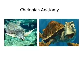
Chelonian Anatomy
200 likes | 635 Views
Chelonian Anatomy. CAUSES TO IMAGE TURTLES. Traumatic injuries of the shell and the ingestion of fishhooks, gravel/stones are the most frequent causes of admission to rescue centers Pulmonary disease can frequently occur secondary to damage to the carapace Dorsal position of the lungs.

Chelonian Anatomy
E N D
Presentation Transcript
CAUSES TO IMAGE TURTLES • Traumatic injuries of the shell and the ingestion of fishhooks, gravel/stones are the most frequent causes of admission to rescue centers • Pulmonary disease can frequently occur secondary to damage to the carapace • Dorsal position of the lungs
AXIAL SKELETON • Composed of the • Carapace • Plastron bones • Vertebrae • Ribs • And derivatives of the ribs
CARAPACE • Carapace is formed by: • Scutes: outermost layer of the shell • Multiple scutes overlap the bony plates that are fused with the vertebrae or ribs forming a single carapace bone • Bony plates: main structural component of the shell • Neural, pleural and peripheral bones
PLASTRON • VENTRAL SURFACE OF THE SHELL • LIMITATIONS OF RADIOGRAPHY IN TURTLES • SUPERIMPOSITIONING OF CARAPACE AND PLASTRON • USEFULNESS OF CT
SKELETAL SYSTEM SCAPULA HUMERUS ACROMION SCAPULA CORACOID
1: ventricle; 2: right atrium; 3 left atrium 4: pectoral musculature 5’ and 5’’ left and right hepatic lobes, respectively; 6: stomach; 7: gallbladder 8: esophagus 9: trachea 10: small intestine 11:large intestine 12: rectum
RESPIRATORY SYSTEM • LUNGS ARE NOT LOBED • PULMONARY PARENCHYMA IS HIGHLY RETICULATED
1 central bronchus 2 pulmonary vasculature 3 pulmonary artery 4 pulmonary vein
1 scapula 2 1st pair of ribs 3 8th cervical vertebra, 4 3rd dorsal vertebra 5 pulmonary parenchyma.
RESPIRATORY PATHOLOGY • Inflammatory exudates particularly those associated with infectious diseases tend to accumulate in the dependent portion of the lung • Pneumonia: seen as loss of normal reticular lung with alveolar pattern, typically seen in the ventral lung
1 kidneys 2 intestinal loop 3 pubic bones. 4 renal veins 5 branch of the external iliac vein 6 aorta artery
RENAL SYSTEM • Kidneys located retroperitoneally between the peritoneum and the shell • Metanephric • Kidney tubules are drained by the ureters(metanephric ducts) and empty into the dorsal cloaca • Lack a distinct cortx, medulla, and renal pelvis • Seen on CT as homogenous parenchyma contrasting only with the perinephric fat between the kidneys and carapace
ARTERIAL SYSTEM • THREE CHAMBERED HEART • 2 ATRIA • 1 VENTRICLE
REFERENCES • A.L.S. Valente a, R. Cuenca a, M. Zamora b, M.L. Parga c, S. Lavin a, F. Alegre c, I. Marco. Computed tomography of the vertebral column and coelomic structures in the normal loggerhead sea turtle (Carettacaretta). The Veterinary Journal 174 (2007) 362–370 • Witherington, Dawn, Wyneken, Jeanette. Chelonian Anatomy. Exotic DVM Vol 4.6. January 2003 • Valente A, CuencavR, Parga M, Lavin S, Franch J, and Marco I. Cervical and coelomic radiologic features of the loggerhead sea turtle, CarettacarettaCan J Vet Res. 2006 October; 70(4): 285–290.