CNS
CNS. Central Nervous System. CNS. Brain and Spinal Cord. CNS: PROTECTION. Turn to page 220-221. CNS: PROTECTION. BONE : Cranium = brain Vertebrate = spine. CNS: PROTECTION. Meninges : Fiborus tissue Color code a,b, b1,c. CNS: PROTECTION. Dura Mater : Toughest connective tissue

CNS
E N D
Presentation Transcript
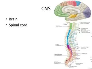
CNS Central Nervous System
CNS • Brain and Spinal Cord
CNS:PROTECTION • Turn to page 220-221
CNS:PROTECTION • BONE: • Cranium = brain • Vertebrate = spine
CNS:PROTECTION • Meninges: Fiborus tissue • Color code a,b, b1,c
CNS:PROTECTION • Dura Mater: Toughest connective tissue • Right under cranium
CNS:PROTECTION • Arachnoid: Spider-web covering under dura mater.
CNS:PROTECTION • Subarachnoid: Pockets of the arachnoid that contain cerebral spinal fluid (CSF)
CNS:PROTECTION • Pia Mater: Delicate, contains many blood vessels.
CNS:PROTECTION • CSF: Circulates subarachnoid space • Cushions, and nourishes. • Surrounds entire CNS
Ohio State University: Neurology: College of Medicine. • CSF from the lumbar region contains 15 to 45 mg/dl protein (lower in childen) and 50-80 mg/dl glucose (two-thirds of blood glucose). Protein concentration in cisternal and ventricular CSF is lower. Normal CSF contains 0-5 mononuclear cells. The CSF pressure, measured at lumbar puncture (LP), is 100-180 mm of H2O (8-15 mm Hg) with the patient lying on the side and 200-300 mm with the patient sitting up.
Problems • Increased protein: In bacterial meningitis, CSF protein may rise to 500 mg/dl. A more moderate increase (150-200 mg/dl) occurs in inflammatory diseases of meninges (meningitis, encephalitis), intracranial tumors, subarachnoid hemorrhage, and cerebral infarction. A more severe increase occurs in the Guillain-Barré syndrome and acoustic and spinal schwannoma.
Problem • Xanthochromia (blonde color) of the CSF following subarachnoid hemorrhage is due to oxyhemoglobin which appears in 4 to 6 hours and bilirubin which appears in two days. Xanthochromia may also be seen with hemorrhagic infarcts, brain tumors, and jaundice.
Normal • Clear as water • Abnormal findings • Faint yellow, orange or pink (Xanthochromia) • CSF Protein >100 mg/dl • Red Blood Cell lysis • Red Blood Cell >100,000/mm3 (Subarachnoid Hemorrhage) • Cloudy or turbid • CSF Leukocytes > 200 wbc/mm3 • Red Blood Cells > 400 per mm3 • Brown or Dark CSF • Metastatic Melanoma (meningeal Melanomatosis) • Jaundice (Hyperbilirubinemia) • Green CSF • Hyperbilirubinemia • Purulent cerebrospinal fluid
(1) intervertebral discs, (2) vertebral bodies, (3) dura, (4) epidural space, (5) spinal cord, and (6) subdural space
BRAIN • Folded to increase surface area • 35 billion neurons (98%) • Adult = 3 lbs
Brain • Brain (3 lbs) at rest needs as much oxygen as 61 lbs of skeletal muscle.
Brain • Turn to page 224
BRAIN • Gyrus: Peaks of the folds, ridges. • Sulcus: furrow or groove between gyrus
Brain • Ventricles: CSF circulate in four major canals. (Travels through brain and into spine) Continuous. • Blue on page 228
BRAINSTEM • Lower brain • Unconscious part
BRAINSTEM • COLOR CODE: • Med. Oblongata = k • Pons = f • Midbrain = a • Reticular formation = g
MEDULLA OBLONGATA • Breathing • Heart rate • Reflex center
PONS • Connects cerebellum to cerebrum • Breathing
Midbrain • Like a hook • Diencephalon to cerebrum • Eye reflexes
Reticular formation • Fibers in the middle of brainstem (connects to RAS) • Inactive so are you! Consciousness.
COLOR • Color code the Diencephalon to the right. • Thalamus = a • Hypothalamus = b
Diencephalon • On top of brainstem
Thalamus • Relay station for sensory headed to the cerebrum. • Filters out messages.
RAS • Reticular Activation System • Deals with arousal and consciousness.
HYPOTHALAMUS • Maintains homeostasis (temp) • Emotions: Rage, pleasure, pain, thirst, hunger
COLOR CODE • On middle picture page 10 • Cerebellum = h • Arbor Vitae = i
CEREBELLUM • Controls muscle balance and coordination. • Lower, posterior part of brain.
Arbor Vitae • White “tree-shaped” structure inside cerebellum.