Download

1 / 29
290 likes | 309 Views
Learn about the supply and transportation of oxygen and nutrients, waste removal, body temperature regulation, and disease protection by the cardiovascular system. Understand the structure and functions of the heart, blood vessels, and blood circulation pathways. Explore heart rate, stroke volume, cardiac output, blood composition, blood vessels, blood pressure, and its regulation during exercise. Discover the importance of maintaining optimal blood pressure levels and how it impacts overall health and fitness.

E N D
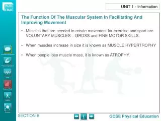
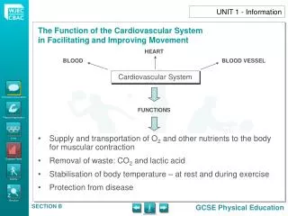
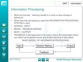
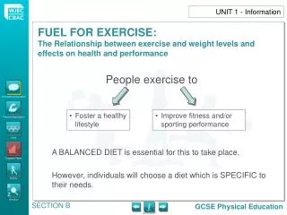
Supply and transportation of O2 and other nutrients to the body for muscular contraction Removal of waste: CO2 and lactic acid Stabilisation of body temperature – at rest and during exercise Protection from disease UNIT 1 - Information HEART BLOOD BLOOD VESSEL Cardiovascular System FUNCTIONS
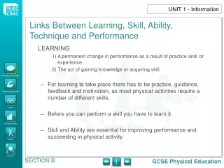
The HEART is a CARDIAC MUSCLE The heart acts as a PUMP in a DOUBLE CIRCULATORY SYSTEM Pulmonary Artery Pulmonary Vein UNIT 1 - Information Structure of the Heart Delivers Carbon Dioxide Capillaries in the lungs Collects Oxygen Aorta L R Blood low in oxygen (deoxygenated) Vena Cava Blood rich in oxygen (oxygenated) Capillaries in the body Aorta Delivers Oxygen and food Collects Carbon Dioxide and waste Show blood circulation
L R UNIT 1 - Information
The RIGH HAND SIDE of the heart pumps DEOXYGENATED BLOOD returning from the body: Deoxygenated blood flows through the VENA CAVA RIGHT ATRIUM RIGHT VENTRICLE PULMONARY ARTERY to the LUNGS for oxygenation Pulmonary artery Deoxygenated blood to the lungs Deoxygenated blood from the body Vena cava Right atrium Tricuspid valve Right ventricle UNIT 1 - Information Show path of deoxygenated blood
Oxygenated blood to the body Aorta Oxygenated blood from the lungs Pulmonary vein Right atrium Bicuspid valve Right ventricle UNIT 1 - Information • The LEFT HAND SIDE of the heart pumps OXYGENATED BLOOD returning from the lungs: • Oxygenated blood through the PULMONARY VEIN LEFT ATRIUM LEFT VENTRICLE AORTA to body. Show path of oxygenated blood
HEART RATE (HR) ‘The number of times the heart beats in one minute.’ At rest it beats between 50 and 80 times per minute. When more blood is required by the muscles during exercise, the heart rate can increase to over 200 beats per minute, pumping around 45 litres around the body. Heart rate varies according to age, fitness and health. UNIT 1 - Information STROKE VOLUME (SV) ‘The amount of blood forced out of the heart (left ventricle) per beat.’
CARDIAC OUTPUT (CO) ‘The amount of blood pumped out of the heart (left ventricle) in one minute.’ Cardiac output varies depending on the intensity of the exercise and the fitness levels of the person. UNIT 1 - Information Cardiac Output Q = Heart Rate × Stroke Volume CO = HR × SV
COMPOSITION OF BLOOD The human body contains nearly 5 litres of blood Blood is made up of 4 elements: UNIT 1 - Information Information/Discussion Practical Application
BLOOD VESSELS Blood is transported from the heart around the body and back to the heart in blood vessels. There are 3 types of blood vessels. UNIT 1 - Information When blood leaves the heart – passes into ARTERIES These branch off into ARTERIOLES – smaller, but more numerous Oxygen diffuses from blood into tissues through thin capillary walls. Carbon dioxide diffuses out of the tissues into the blood When it reaches the muscles, blood passes into CAPILLARIES – even smaller, but more numerous. Deoxygenated blood Oxygenated blood At the capillaries, the blood gives up its oxygen and takes in carbon dioxide The blood starts its journey BACK to the heart in small, narrow veins called VENULES Artery Vein Arterioles Venules Capillaries The blood then passes into larger VEINS before returning to the heart
BLOOD PRESSURE The force of blood against the artery walls. With exercise, the heart has to work to supply more O2 to muscles. As a result, the force of blood leaving the heart increases and blood pressure increases. Blood pressure is easily measured by taking the pressure at an artery in the arm. SYSTOLIC PRESSURE is the pressure of blood flow on the arteries when the LEFT VENTRICLE CONTRACTS. DIASTOLIC PRESSURE is the pressure of blood flow on the arteries when the LEFT VENTRICLE RELAXES. UNIT 1 - Information
BLOOD PRESSURE The average blood pressure reading for a young adult is 120/80. Factors which can affect blood pressure: AGE, SEX, EXERCISE INTENSITY, STRESS, CIRCULATORY SYSTEM, FITNESS Ways in which blood pressure can be reduced: REGULAR EXERCISE, SENSIBLE DIET, AVOID STRESS, MEDICATION The blood flow and blood distribution change according to the demand of exercising. The working parts of the body need to be supplied with the necessary amounts of O2 The re-distribution of blood is called the VASCULAR SHUNT UNIT 1 - Information
BLOOD PRESSURE Blood flow to the muscles and the skin will increase during exercise. Blood flow to the kidneys and digestive system will decrease during exercise. The heat produced by the body increases as the INTENSITY and DURATION of exercise increases. To control high body temperature, blood is diverted to the capillaries just below the skin – this causes the skin to redden and heat from the blood is then RADIATED from the skin. This widening of the capillaries is called VASODILATION. To control low body temperature, the capillaries VASO CONSTRICT – become narrower, therefore reducing heat loss by radiation. Muscles begin to ‘shiver’ – small contractions which provide ‘heat’. UNIT 1 - Information
BLOOD PRESSURE Another way of combating overheating is by SWEATING. Sweat is formed in sweat glands under the skin. Sweating is caused by the EVAPORATION of sweat from the skin’s surface. UNIT 1 - Information
Group Discussion Heart / Blood / Blood Vessels FUNCTIONS Mapping exercise Double Loop System – Transportation of Blood (Blood Pathway) Discussion ‘As a result of regular Aerobic Training, the heart gets bigger (HYPERTROPHY). How does this effect : Stroke volume, Heart rate, Blood flow Cardiac output UNIT 1 – Practical Application
Group Discussion Heart rate 1) At rest, 2) after exercise, 3) Recovery rate (2mins/15mins) Discussion ‘How would heart rate differ between a short period of anaerobic work and a longer period of aerobic work?’ UNIT 1 – Practical Application
Use the following 2 diagrams to relate heart rate to physical activity: 1) The graph below shows the heart rate of two sixteen year old athletes when training at the same intensity. Explain why athlete B is the fittest athlete. 180 Athlete A Athlete B 120 Heart Rate (beats per minute) 90 60 0 20 10 30 Time (minutes) UNIT 1 – Practical Application
2) The graph below shows the heart rate of an eighteen year old badminton player during a game. Heart Rate (beats per minute) 250 200 150 100 50 10 15 20 5 0 Time (minutes) UNIT 1 – Practical Application • Give two pieces of evidence to suggest that this player is a fit competitor.
Heart Rate (beats per minute) 250 200 150 100 50 10 15 20 5 0 Time (minutes) UNIT 1 – Practical Application • During the game the player’s heart rate reaches 220 beats per minute (BPM). Calculate the player’s maximum heart rate (MHR) during the game. • What evidence is there to suggest that this player worked both aerobically and anaerobically during the game?
Skeletal System Respiratory System Aerobic / Anaerobic Systems Muscular System Energy Continuum Training Zones Intensity / Duration of Exercise Short-term effects of exercise on the systems of the body Long-term effects of exercise on the systems of the body UNIT 1 - Links
UNIT 1 - Activity • Match the parts of the heart and connecting blood vessels to their function. [Click here to see diagram] Left ventricle Tricuspid valve Pulmonary vein Aorta Superior vena cava Left atrium Right atrium Inferior vena cava Bicuspid valve Right ventricle Pulmonary artery
Deoxygenated blood to the lungs Oxygenated blood to the body Pulmonary artery Aorta Deoxygenated blood from the body Oxygenated blood from the lungs Pulmonary vein Vena cava Left atrium Right atrium Bicuspid valve Tricuspid valve Left ventricle Right ventricle UNIT 1 - Activity SHOW/HIDE LABELS Back SHOW/HIDE ARROWS
UNIT 1 - Activity • Explain the relationship between cardiac output (Q) and exercise intensity. • Explain how the heart’s structure is adapted to its function. • What is blood pressure? • What is systolic pressure? • What is diastolic pressure? • What is the normal blood pressure reading for a young person? • Give five factors that can affect blood pressure.
UNIT 1 - Activity • Why can narrowing or blocking of blood vessels be dangerous? • Give five ways blood pressure can be reduced. • Define the following terms: • Heart rate (HR) • Stroke volume (SV) • Cardiac output (Q) • What simple equation relates these three values? • Give two differences between cardiac and skeletal muscle.
UNIT 1 - Activity • Complete the following description of the blood’s journey from the heart around the body and back to the heart by dragging the correct word from the list below: Blood is transported from the heart around the __________ and back to the heart in ______________________. There are ___________ types of blood vessel. These branch off into ________________. These are smaller but are more numerous. When it gets to the muscles, blood passes into the ___________. These are even smaller, but there are millions of them. At the capillaries, the blood gives up its _______________ and takes in _______________. The blood starts its journey back to the heart in small veins called ________________. The blood then passes into larger _______________ before returning to the heart. venules capillaries arteries blood vessels body oxygen arterioles veins carbon dioxide three
UNIT 1 - Activity • Explain four ways in which blood helps the body during exercise. • Complete the table to show how the constituents (parts) of blood help us when doing sport. • The path that the blood takes can be described as a double loop. What is each loop called?
UNIT 1 - Activity • How would a 1500m runner benefit from higher levels of red blood cells? • Complete the following table:
UNIT 1 - Activity • X on the graph shows how a sports person’s heart rate responds to a 10 minute run at 12kmph and how it recovers. Y shows the heart rate response to the same run after a period of regular endurance training. Explain the reasons for the changed heart rate pattern. BPM 180 170 160 150 140 130 120 110 100 90 80 70 X Y TIME 0 mins 10 mins 20 mins
UNIT 1 - Activity • Answer the following: • How does regular aerobic training affect stroke volume? • How does this affect a person’s heart rate and cardiac output when running at a medium pace for 5 minutes?
Healthy Lifestyles HEART Blood Vessels Blood FUNCTIONS UNIT 1 – Key Facts/ Glossary Systemic • Double circuit Pulmonary • Blood flow • Cooling of body • Cardiac Output (CO) • Composition of blood Vasodilatation (Sweating) Vasoconstriction (Low body temperature control) • Arteries / Veins / Capillaries • Gaseous Exchange • Blood Pressure (BP)