Download
1 / 53
650 likes | 1.07k Views
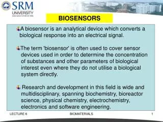
Introduction to biosensors. Peter Bienstman. Biosensors. Detect presence and concentration of biomolecules DNA Proteins Virus Bacteria … Two classes: Labeled: indirect detection Label-free: direct detection. Applications. Diagnostics Drug development Food safety

E N D
Introduction to biosensors Peter Bienstman
Biosensors • Detect presence and concentration of biomolecules • DNA • Proteins • Virus • Bacteria • … • Two classes: • Labeled: indirect detection • Label-free: direct detection
Applications • Diagnostics • Drug development • Food safety • Environmental monitoring • …
Desired characteristics • Low limit of detection (“sensitivity”) • Selective • Reproducible • Cheap • Portable • Fast • Multi-parameter • …
Labeled optical sensor types • Many, many types • E.g. • Elisa • Au nanoparticle labels • Quantum dot labels • Bead-based assays • Padlock probes • Not an exhaustive list!
Elisa tests • Enzyme-LinkedImmunoSorbentAssay • Workhorse of protein detection • Detect protein by using • fluorescent labels • labels with enzymes that start a colouring reaction on a dye substrate • …
Example: pregnancy test • Detects hCG protein (human Chorionic Gonadotropin) in urine • Based on strip which pulls fluid through by capillary action (lateral flow immunochromatography)
Test principle See animations at http://www.whfreeman.com/kuby/content/anm/kb07an01.htm
Assay zones Fluid flows through 3 zones: • R: reaction zone: hCG picks up free antibody labeled with enzyme • T: test zone: hCG+antibody+enzyme gets bound by immobilised antibody on strip, enzyme starts colouring reaction of dye if pregnant • C: control zone: antibody picks up any remaining antibody+enzyme complexes, enzyme starts colouring if test works OK
Variations of pregnancy test • Don’t use enzymes to colour a dye, but use gold nanoparticles • About 10 nm in diameter • Au is nice because it’s easy to functionalise it • Red in colour, but depends on particle size (see later)
Au nanoparticles Immobilised on latex beads Two different particles sizes In solution
Ways to use them • As a fancy dye • Changing colour on aggregation • Combined with latex beads • …
As fancy dye • Just use them as a dye, i.e. instead of the enzyme • If there are enough of them in the test zone, they will give a red line • Used e.g. by UltiMed pregnancy test
Changing colour on aggregation Colloidal gold coated with hCG antibody
Changing colour on aggregation hCG present
Changing colour on aggregation Absorption band shifts due to aggregation and colourchanges (see later)
Combined with latex beads • Au nanoparticles and latex microparticles • When pregnant, Au colours the latex bead and a size filter prevents them from washing downstream
Quantum dot labels • Alternative to metallic nanoparticles • Typically colloidally grown • PbSe, CdTe, … • Much sharper spectra, widely tuneable by size
Multiparameter assays • Pregnancy test measures only single compound • Very interesting to have more than 1 target • Multiplexed, multi-parameter assays • Two formats: • 2D arrays on chip: spatial encoding • Free floating labeled microcarriers
Labeled microcarriers • Don’t flow fluid over planar substrate, but break up substrate into microcarriers which float in the fluid • Better mixing properties too
Read-out in flow cytometer • E.g., one laser measures label on bead, the other measures the reporter fluorophore
Colour-encoded beads e.g. LuminexxMAP technology, 2 fluorescent dyes in different ratios
Labeling • detect a molecule by attaching a label to it • very sensitive (10-9...10-16mol/l) • commercial product (Elisa, DNA arrays, ..)
Disadvantages to labeling • some labels are very costly • only measures final state, no kinetics • label can influence properties of biomolecules • strong interest in label-free sensors
Label-free sensors • detect presence of biomolecules directly • focus here: label-free optical biosensors • selective binding causes refractive index change flow with biomolecules matching biomolecule (analyte) biorecognition element (ligand)
Index change • How to measure the refractive index change? • Surface plasmon sensors • Evanescent wave sensors • Mach-Zehnder interferometer • Resonant cavities • Once again, the list is not exhaustive. • Also, there are many non-optical techniques (impedimetric, mass, …)
Plasmons • Collective wave oscillations of electrons in a metal motion of electrons propagation of wave Fig: R. Nave, Hyperphysics
Surface plasmons • Interaction between: • plasmon at surface of metal • electromagnetic wave EM wave plasmon
Magnitude of EM field Cannot be excited directly from the outside position light intensity
angle Reflection experiment reflection angle
Towards a biosensor reflection angle angle
R From source To detector Prism Gold Surface plasmon resonance • Popular for biosensing (Biacore machine) • High fields near the interface are very sensitive to refractive index changes • Gold is very suitable for biochemistry
advantages • very sensitive, index differences of 10-6 possible • functionalised Au layers off-the-shelf available • integrated microfluidics • but • bulky • expensive • difficult to integrate and multiplex
Evanescent wave biosensor Densmore, 2008
Influence of mode profile • profile should overlap maximally with the adlayer, and not with bulk fluid (noise!) • high index contrast is best Low contrast High contrast
Effective index change still needs to be translated into something measurable. • Many possibilities: • Resonators • Interferometers • …
Ring resonators 1.55 μm Binding of biomoleculeschange of refractive index resonance wavelength shift
Towards a better sensor • High demandsonread-out system, but filters noise Larger shift Narrower dips More interactionbetweenlight and molecules
Sensitivity vs detection limit • Sensitivity: shift of resonance wavelength (in nm) for a given excitation, e.g. • Bulk sensitivity: nm / RIU (refractive index unit) • Adlayer sensitivity: nm / nm • Detection limit: smallest measurable excitation Δλmin : smallest distinguishablewavelength shift
What determines Δλmin ? • precision of measurement equipment • noise in the system (thermal, mechanical, …) • design of the sensor • e.g.: higher Q is better • often in conflict with sensitivity • quality of data analysis • averaging • analytical curve fitting • Δλmin can get smaller than measurement resolution!