Chapter 20 - Enterobacteriaceae
Chapter 20 - Enterics. Family Enterobacteriaceae often referred to as enterics"Four major features:All ferment glucose (dextrose)All reduce nitrates to nitritesAll are oxidase negative, except PlesiomonasAll except Klebsiella, Shigella and Yersinia are motile. Microscopic and Colony Morphology

Chapter 20 - Enterobacteriaceae
E N D
Presentation Transcript
1. Chapter 20 - Enterobacteriaceae MLAB 2434 � Clinical Microbiology
Keri Brophy-Martinez
2. Chapter 20 - Enterics Family Enterobacteriaceae often referred to as �enterics�
Four major features:
All ferment glucose (dextrose)
All reduce nitrates to nitrites
All are oxidase negative, except Plesiomonas
All except Klebsiella, Shigella and Yersinia are motile
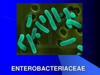
3. Microscopic and Colony Morphology Gram negative bacilli or coccobacilli
Non-spore forming
Facultative anaerobe
Colony morphology on BAP or CA of little value, as they look the same( large, moist and grey on SBA)
Selective and differential media are used for initial colony evaluation (ex. MacConkey, HE, XLD agars)
4. Classification of Enterics Due to the very large number of organisms in the Family Enterobacteriaceae (see Table 20-1, page 505), species are grouped into Tribes, which have similar characteristics (Table 20-2, page 506)
Within each Tribe, species are further subgrouped under genera
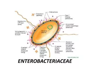
5. Virulence and Antigenic Factors of Enterics Ability to colonize, adhere, produce various toxins and invade tissues
Some possess plasmids that may mediate resistance to antibiotics
ESBL�s: Extended-Spectrum beta lactamases
Inactivate extended spectrum antibiotics like penicillins, cephalosporins and aztreonam
6. Virulence and Antigenic Factors of Enterics Many enterics possess antigens that can be used to identify groups
O antigen � somatic, heat-stable antigen located in the cell wall, made by all bacteria, stimulates EARLY antibody production
H antigen � flagellar, made by bacteria with flagella, heat labile antigen,stimulates LATE antibody production
K antigen � capsular, heat-labile antigen, made by some bacteria
Vi- capsular antigen of S. typhii
7. Clinical Significance of Enterics Enterics are ubiquitous in nature
Except for few, most are present in the intestinal tract of animals and humans as commensal flora; therefore, they are sometimes call �fecal coliforms�
Some live in water, soil and sewage
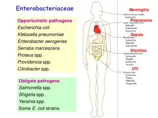
8. Clinical Significance of Enterics (cont�d) Based on clinical infections produced, enterics are divided into two categories:
Opportunistic pathogens � normally part of the usual intestinal flora that may produce infection outside the intestine
Primary intestinal pathogens � not commensal flora, they produce infection from ingestion of contaminated food or water (Salmonella, Shigella, and Yersinia sp.)
9. Escherichia coli Most significant species in the genus
Important potential pathogen in humans
Common isolate from colon flora
10. Escherichia coli (cont�d) Characteristics
Dry, pink (lactose positive) colony with surrounding pink area on MacConkey
11. Escherichia coli (cont�d) Ferments glucose, lactose, trehalose, & xylose
Positive indole and methyl red tests
Does NOT produce H2S or phenylalanine deaminase
Simmons citrate negative
Usually motile
Voges-Proskauer test negative
Fimbriae
O1, H, K antigens
12. Escherichia coli (cont�d) Infections
Wide range including meningitis, gastrointestinal, urinary tract, wound, and bacteremia
Gastrointestinal Infections
Enteropathogenic (EPEC) � primarily in infants and children; outbreaks in hospital nurseries and day care centers; stool has mucous but not blood; identified by serotyping
13. Escherichia coli (cont�d) Enterotoxigenic (ETEC) � �traveler�s diarrhea�; watery diarrhea without blood; self-limiting; usually not identified, other than patient history and lactose-positive organisms cultured on differential media
Enteroinvasive (EIEC) � produce dysentery with bowel penetration, invasion and destruction of intestinal mucosa; watery diarrhea with blood; do NOT ferment lactose; identified via DNA probes
14. Escherichia coli (cont�d) Enterohemorrhagic (EHEC serotype 0157:H7)
Origin
Isolated in 1970
Undercooked hamburger, unpasteurized milk and apple cider have spread the infection
Pathogenesis
Circulating Shiga toxin binds to kidney endothelium causing an inflammatory response
Macrophages and neutrophils damage the endothelium and glomerular basement membrane
15. Escherichia coli (cont�d) Enterohemorrhagic (EHEC serotype 0157:H7)
Clinical Symptoms
Starts with a watery diarrhea then progresses to bloody diarrhea. No WBC�s are found in stool
Associated with hemorrhagic diarrhea, TTP and hemolytic-uremic syndrome (HUS), which includes low platelet count, hemolytic anemia, and kidney failure; potentially fatal, especially in young children;
Laboratory Diagnosis
Does NOT ferment sorbitol
Identified by serotyping, latex tests
16. Escherichia coli (cont�d) Enteroaggregative (EaggEC/EAEC) � cause diarrhea by adhering to the mucosal surface of the intestine; watery diarrhea; symptoms may persist for over two weeks
Enteroadherent
Diffusely adherent(DAEC), associated with UTI�s and diarrheal disease, esp. in children and pregnant women
Urinary Tract Infections
E. coli is most common cause of UTI and kidney infection in humans
Usually originate in the large instestine
Able to adhere to epithelial cells in the urinary tract
17. Escherichia coli (cont�d) Septicemia & Meningitis
E. coli is one of the most common causes of septicemia and meningitis among neonates; acquired in the birth canal before or during delivery
E. coli also causes bacteremia in adults, primarily from a genitourinary tract infection or a gastrointestinal source
Escherichia hermannii � yellow pigmented; isolated from CSF, wounds and blood
Escherichia vulneris - yellow pigmented; wounds
18. Klebsiella, Enterobacter, Serratia & Hafnia sp. Usually found in intestinal tract
Wide variety of infections, primarily pneumonia, wound, and UTI
General characteristics:
Some species are non-motile
Simmons citrate positive
H2S negative
Phenylalanine deaminase negative
Some weakly urease positive
MR negative; VP positive
19. Klebsiella species Usually found in GI tract
Four major species(Table 20-5, page 513)
K. pneumoniae is mostly commonly isolated species
Possesses a polysaccharide capsule, which protects against phagocytosis and antibiotics AND makes the colonies moist and mucoid
Has a distinctive �yeasty� odor
Frequent cause of nosocomial pneumonia
20. Klebsiella species (cont�d) Significant biochemical reactions
Lactose positive
Most are urease positive
Non-motile
21. Enterobacter species Comprised of 12 species; E. cloacae and E. aerogenes are most common
Isolated from wounds, urine, blood and CSF
Major characteristics
Colonies resemble Klebsiella
Motile
Simmons citrate positive
MR negative; VP positive
22. Enterobacter species (cont�d)
23. Serratia species Seven species, but S. marcescens is the only one clinically important
Frequently found in nosocomial infections of urinary or respiratory tracts
Implicated in bacteremic outbreaks in nurseries, cardiac surgery, and burn units
Fairly resistant to antibiotics
24. Serratia species (cont�d) Major characteristics
Ferments lactose slowly
Produce characteristic pink or red pigment, especially when cultures are left at room temperature
S. marscens on
nutrient agar ?
25. Hafnia species Hafnia alvei is only species
Has been isolated from many anatomical sites in humans and the environment
Occasionally isolated from stools
Delayed citrate reaction is major characteristic
26. Proteus, Morganella & Providencia species All are normal intestinal flora
Opportunistic pathogens
Deaminate phenylalanine
All are lactose negative
27. Proteus Lactose negative
28. Proteus species P. mirabilis and P. vulgaris are widely recognized human pathogens
Isolated from urine, wounds, and ear and bacteremic infections
Both produce swarming colonies on non-selective media and have a distinctive �burned chocolate� odor
Both are strongly urease positive
Both are phenylalanine deaminase positive
29. Proteus species (cont�d) A exhibits characteristic �swarming�
B shows urease positive on right
30. Morganella species Morganella morganii is only species
Documented cause of UTI
Isolated from other anatomical sites
Urease positive
Phenylalanine deaminase positive
31. Providencia species Providencia rettgeri is pathogen of urinary tract and has caused nosocomial outbreaks
Providenicia stuartii can cause nosocomial outbreaks in burn units and has been isolated from urine
Both are phenylalanine deaminase positive
32. Citrobacter species Citrobacter freundii associated with nosocomial infections (UTI, pneumonias, and intraabdominal abscesses)
Ferments lactose and hydrolyzes urea slowly
Methyl red positive, Simmons citrate positive
Resembles Salmonella sp.
33. Salmonella Produce significant infections in humans and certain animals
On differential selective agar, produces clear, colorless, non-lactose fermenting colonies with black centers (if media contains indicator for hydrogen sulfide)
34. Salmonella (cont�d) Salmonella on MacConkey
35. Salmonella (cont�d) Lactose negative
Negative for indole, VP, phenylalanine deaminase, and urease
Most produce H2S
Do not grow in potassium cyanide
36. Salmonella Virulence Factors
Fimbriae- help in attachment
Transverse intestinal mucosa (not known how)
Enterotoxin
Anitgenic Structures
O and H structures are used for serologic grouping
O- heat stable, lipopolysaccharide on the outer membrane of cell wall
H- heat labile
Vi- helpful in ID of Salmonella typhi
Heat labile, surface polysaccharide capsular antigen
37. Salmonella (cont�d) Clinical Infections
Acute gastroenteritis or food poisoning
Source = handling pets, insufficiently cooked eggs and chicken,milk, and contaminated cooking utensils
Occurs 8 to 36 hours after ingestion
Symptoms include vomiting, chills, watery diarrhea and abdominal pain
Requires a high microbial load for infection
Self-limiting in healthy individuals (antibiotics and antidiarrheal agents may prolong symptoms)
38. Salmonella (cont�d) Typhoid and Other Enteric Fevers
Prolonged fever
Bacteremia
Involvement of the RE system, particularly liver, spleen, intestines, and mesentery
Dissemination to multiple organs
Occurs more often in tropical and subtropical countries
39. Salmonella (cont�d) Typhoid fever
S. Typhii causative organism
Invades intestinal mucosa causing constipation
Gains entrance into lymphatic system and bloodstream then into the liver, spleen and bone marrow where they are phagocytized by PMN�s. They multiply in the PMN�s and are eventually released into the blood stream.
Finally, they invade the gall bladder and other parts of the intestinal tract to initiate GI symptoms.
Found in contaminated food originating from infected individuals
Other causes, improper disposal of sewage, poor sanitation, lack of modern H2O systems
Develops 9-14 days after ingestion with symptoms of fever, malaise, anorexia, myalgia, continuous dull headache
Severe
40. Salmonella (cont�d) Salmonella Bacteremia
Carrier State
Organisms shed in feces
Gallbladder is the site of organisms (removal of gallbladder may be the only solution to carrier state)
41. Shigella species Closely related to the Escherichia
All species cause bacillary dysentery, not normal GI flora
S. dysenteriae (Group A)
S. flexneri (Group B)
S. boydii (Group C)
S. sonnei (Group D)
42. Shigella (cont�d) Characteristics
Non-motile, lack the H antigen
Do not produce gas from glucose
Do not hydrolyze urea
Do not produce H2S on TSI
On differential, selective media appear as clear, NLF
Lysine decarboxylase negative
ONPG positive (delayed lactose +)
Fragile organisms
Possess O and some have K antigens
43. Shigella (cont�d) Clinical Infections
Cause dysentery (bloody stools, mucous, and numerous WBC)
Confined to GI tract
Septicemia rare
Humans are only known reservoir
Oral-fecal transmission, person-person
Fewer than 200 bacilli are needed for infection in healthy individuals= takes a low infective dose
44. Shigella (cont�d)
45. Yersinia species Consists of 11 named species
Yersinia pestis
Causes plague, which is a disease primarily of rodents; transmitted by rat fleas
Two forms of plague
bubonic- flea bite, buboes
Pneumonic- secondary to bubonic, organisms multiply in bloodstream and resp. tract
Gram-negative, short, plump bacillus, exhibiting �safety-pin� or �bipolar� staining
46. Characteristic bubo
47. Yersinia gram stain
48. Yersinia species Yersinia enterocolitica
Most common form of Yersinia
Found worldwide
Found in pigs, cats and dogs
Human also infected by ingestion of contaminated food or water
Some infections result from eating contaminated market meat and vacuum-packed beef
Is able to survive refrigerator temperatures (can use �cold enrichment� to isolate)
Mainly causes acute gastroenteritis with fever, headaches
Stool may be bloody
Can mimic appendicitis
49. Laboratory Diagnosis of Enterics Collection and Handling
If not processed quickly, should be collected and transported in Cary-Blair, Amies, or Stuart media
Isolation and Identification
Site of origin must be considered
Enterics from sterile body sites are highly significant
Routinely cultured from stool
50. Laboratory Diagnosis of Enterics (cont�d) Media for Isolation and Identification of Enterics
Most labs use BAP, CA and a selective/differential medium such as MacConkey
On BA, produce large, greyish, smooth colonies; can be �-hemolytic or nonhemolytic
On MacConkey, lactose positive are pink; lactose negative are clear and colorless
51. Laboratory Diagnosis of Enterics (cont�d) For stools, highly selective media, such as Hektoen Enteric (HE), XLD, or SS is used along with MacConkey agar
CIN- selects for Yersenia, hold at room temp.
Identification
Most labs use a miniaturized or automated commercial identification system, rather than multiple tubes inoculated manually
52. Laboratory Diagnosis of Enterics (cont�d) Identification (cont�d)
All enterics are
Oxidase negative
Ferment glucose
Reduce nitrates to nitrites
53. Laboratory Diagnosis of Enterics (cont�d) Common Biochemical Tests
Lactose fermentation and utilization of carbohydrates
Triple Sugar Iron (TSI)
ONPG
Glucose metabolism
Methyl red
Voges-Proskauer
54. Laboratory Diagnosis of Enterics (cont�d) Common Biochemical Tests (cont�d)
Miscellaneous Reactions
Indole
Citrate utilization
Urease production
Motility
Phenylalanine deaminase
Decarboxylase tests
55. Identification Clues Lactose Fermenter
Think: E. coli, Klebsiella, Enterobacter, possibly Citrobacter
Lactose Fermenter with mucoid colony
Think: Klebsiella, Enterobacter
H2S +
Think: Proteus, Salmonella, Citrobacter
Nonmotile
Think: Klebsiella, Shigella
Voges- Proskauer +
Think: Klebsiella, Enterobacter, Serratia
56. Screening Stools for Pathogens Because stools have numerous microbial flora, efficient screening methods must be used to recover any pathogens
Enteric pathogens include Salmonella, Shigella, Aeromonas, Campylobacter, Yersinia, Vibrio, and E. coli 0157:H7
57. Screening Stools for Pathogens (cont�d) Most labs screen for Salmonella, Shigella, and Campylobacter; many screen for E. coli 0157:H7
Fecal pathogens are generally lactose-negative (although Proteus, Providencia, Serratia, Citrobacter and Pseudomonas are also lactose-negative)