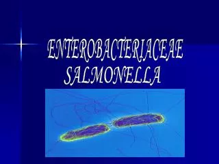
ENTEROBACTERIACEAE
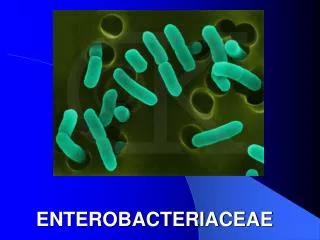
ENTEROBACTERIACEAE. Morphology & Identification. Gram-negative non-spore forming rods. When motile, by peritrichous flagella. Primarily normal flora of gastrointestinal tract. E. coli > Klebsiella > Proteus > Enterobacter Free living, also transient colonizers of skin.

ENTEROBACTERIACEAE
E N D
Presentation Transcript
Morphology & Identification • Gram-negative non-spore forming rods. When motile, by peritrichous flagella. • Primarily normal flora of gastrointestinal tract. E. coli>Klebsiella>Proteus>Enterobacter • Free living, also transient colonizers of skin. • Facultative anaerobes: mixed acid fermentation • All ferment glucose; all reduce nitrates to nitrites; all oxidase negative. • Lactose fermentation: normal flora positive and pathogens negative. • Primary isolation media include eosin-methylene-blue (EMB) and MacConkey agar. • Differential selective media for specific organisms including dyes and bile salts. (Salmonella-Shigella (SS) medium, bismuth sulfite media.)
Escherichia Shigella Edwardsiella Salmonella Citrobacter Klebsiella Enterobacter Hafnia Serratia Proteus Providencia Morganella Yersinia Erwinia Pectinobacterium Classification~29 genera, over 100 species.
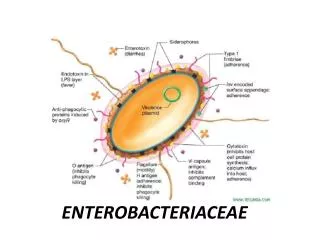
Antigenic Structure • Most are motile by peritrichous flagella --H antigens. • Capsule – Kantigen (Vifor Salmonella). • Cell envelope (wall) • LPS (endotoxin) –O antigen. • various outer membrane proteins. • Pili - various antigen types, some encoded by plasmids
鞭毛抗原(H) K或Vi抗原 菌体抗原(O)
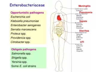
Opportunistic diseases -Enterobacteriaceae • septicemia, • pneumonia, • meningitis • urinary tract infections CitrobacterEnterobacter Escherichia Hafnia Morganella Providencia Serratia
Enterobacteriaceae:gastrointestinal diseases • Escherichia coli • Salmonella • Shigella • Yersinia entercolitica
Reiter's syndrome • Histocompatibility antigen (HLA) B27 • Enterobacteriaceae • Salmonella • Shigella • Yersinia • NotEnterobacteriaceae • Campylobacter • Chlamydia
Enterobacteriaceae • community acquired • otherwise healthy people • Klebsiella pneumoniae • respiratory diseases • prominent capsule • urinary tract infection • fecal contamination • E. coli • Proteus • urease (degrades urea) • alkaline urine
Enterobacteriaceae • gram negative facultative anaerobic rods • – oxidase negative (no cytochrome oxidase)
Feces • E. coli • lactose positive • not usually identified • lactose positive sp. common, healthy intestine • Shigella, Salmonella,Yersinia • lactose negative • identified
Enterobacteriaceae • other sites • identified biochemically
Serotypes • reference laboratory • antigens • O (lipopolysaccharide) • H (flagellar) • K (capsular)
Escherichia coli • Toxins: two types of enterotoxin; Shiga-type toxin; Enteroaggregative ST-like toxin; Hemolysins; Endotoxin • Type III secretion system • Adhesions –colonization factors ; both pili or fimbriae ;non-fimbrial factors involved in attachment. There are at least 21 different types of adhesions. • Virulence factors that protect the bacteria from host defenses: Capsule/Iron capturing ability (enterochelin) • Outer membrane proteins
E. coli fimbriae Type 1 mannose P • galactose • glycolipids • glycoproteins
E.coli-urinary tract infection Is the leading cause of urinary tract infections which can lead to acute cystitis (bladder infection) and pyelonephritis (kidney infection).
E.coli-Meningitis and Sepsis • Neonatal meningitis – is the leading cause of neonatal meningitis and septicemia with a high mortality rate. Usually caused by strains with the K1 capsular antigen.
Enteropathogenic E. coli • fever • infantdiarrhea • vomiting • nausea • non-bloody stools • Destruction of surface microvilli • loose attachment mediated by bundle forming pili (Bfp); • Stimulation of intracellular calcium level; • rearrangement of intracellular actin,
Enterotoxigenic E. coli • A watery diarrhea, nausea, abdominal cramps and low-grade fever for 1-5 days. • Travellers diarrhea and diarrhea in children in developing countries • Transmission is via contaminated food or water.
Enterotoxigenic E. coli • diarrhea like cholera • milder • nursery travellers diarrhea • caused by LT, ST, or LT/ST.
Enterotoxigenic E. coli • Heat labile toxin • like choleragen • Adenyl cyclase activated • cyclic AMP • secretion water/ions • Heat stable toxin • Guanylate cyclase activated • cyclic GMP • uptake water/ions
E.coli-Enteroinvasive (EIEC) • The organism attaches to the intestinal mucosa via pili • Outermembrane proteins are involved in direct penetration, invasion of the intestinal cells, and destruction of the intestinal mucosa. • There is lateral movement of the organism from one cell to adjacent cells. • Symptoms include fever,severe abdominal cramps, malaise, and watery diarrhea followed by scanty stools containing blood, mucous, and pus. • resembles shigellosis
Enteroinvasive E. coli (EIEC) • Dysentery • resembles shigellosis • elder children and adult diarrhea
E.coli-c. Enteropathogenic (EPEC) • Malaiseand low grade feverdiarrhea, vomiting, nausea, non-bloody stools • Bundle forming pili are involved in attachment to the intestinal mucosa. • This leads to changes in signal transduction in the cells, effacement of the microvilli, and to intimate attachment via a non-fimbrial adhesion called intimin. • This is a problem mainly in hospitalized infants and in day care centers.
E.coli-d. Enterohemorrhagic (EHEC) • Hemorrhagic • bloody, copious diarrhea • few leukocytes • afebrile • hemolytic-uremic syndrome • hemolytic anemia • thrombocytopenia (low platelets) • kidney failure
Transmission electron micrograph Enterohemorrhagic E. coli • Usually O157:H7
Enterohemorrhagic E. coli • Vero toxin • “shiga-like” • Hemolysins • younger than 5 years old,causing hemorrhagic colitis
Enteroaggregative E. coli 肠集聚型大肠杆菌 • a cause of persistent, watery diarrhea with vomiting and dehydration in infants. • That is autoagglutination in a ‘stacked brick’ arrangement. • the bacteria adheres to the intestinal mucosa and elaborates enterotoxins (enteroaggregative heat-stable toxin, EAST). • The result is mucosal damage, secretion of large amounts of mucus, and a secretory diarrhea.
E.coli-Enteroaggregative (EAggEC) • Mucous associated autoagglutinins cause aggregation of the bacteria at the cell surface and result in the formation of a mucous biofilm. • The organisms attach via pili and liberate a cytotoxin distinct from, but similar to the ST and LT enterotoxins liberated by ETEC. • Symptoms incluse watery diarrhea, vomiting, dehydration and occasional abdominal pain.
Sanitary significance • Totoal bacterial number: number of bacteria contained per ml or gm of the sample; the standard of drinking water is less than 100. • Coliform bacteria index: the number of coliform bacteria detected out per 1000 ml sample; the standard of drinking water is less than 3
Escherichia coli • Genetically E. coli and Shigella are genetically highly closely related. For practical reasons (primarily to avoid confusion) they are not placed in the same genus. Not surprisingly there is a lot of overlap between diseases caused by the two organisms. • 1) Enteropathogenic E. coli (EPEC). Certain serotypes are commonly found associated with infant diarrhea. The use of gene probes has confirmed these strains as different from other groups listed below. There is a characteristic morphological lesion with destruction of microvilli without invasion of the organism that suggests adhesion is important. Clinically one observes fever, diarrhea, vomiting and nausea usually with non-bloody stools. • 2) Enterotoxigenic E. coli (ETEC) produce diarrhea resembling cholera but much milder in degree. Also cause "traveler’s diarrhea". Two types of plasmid-encoded toxins are produced. a) Heat labile toxins which are similar to choleragen (see cholera section below). Adenyl cyclase is activated with production of cyclic AMP and increased secretion of water and ions. b) Heat stable toxins; guanylate cyclase is activated which inhibits ionic and water uptake from the gut lumen. Watery diarrhea, fever and nausea result in both cases. • 3) Enteroinvasive E. coli (EIEC) produce dysentery (indistinguishable clinically from shigellosis, see bacillary dysentery below). • 4) Enterohemorrhagic E. coli (EHEC). These are usually serotype O157: H7. These organisms can produce a hemorrhagic colitis (characterized by bloody and copious diarrhea with few leukocytes in afebrile patients). Outbreaks are often caused by contaminated hamburger meat. The organisms can disseminate into the bloodstream producing systemic hemolytic-uremic syndrome (hemolytic anemia, thrombocytopenia and kidney failure). Production of Vero toxin (biochemically similar to shiga toxin thus also known as "shiga-like") is highly associated with this group of organisms; encoded by a phage. Hemolysins (plasmid encoded) are also important in pathogenesis. • As noted above, there are at least 4 etiologically distinct diseases. However, in the diagnostic laboratory generally the groups are not differentiated and treatment would be on symptomatology. Generally fluid replacement is the primary treatment. Antibiotics are generally not used except in severe disease or disease that has progressed to a systemic stage (e.g.hemolytic-uremia syndrome). Two major classes of pili are produced by E. coli : mannose sensitive and mannose resistant pili. The former bind to mannose containing glyocoproteins and the latter to cerebrosides on the host epithelium allowing attachment. This aids in colonization by E. coli.
Shigella • S. flexneri, S. boydii, S. sonnei, S. dysenteriae • bacillary dysentery • shigellosis • bloody feces • intestinal pain • pus
Genral features • Pili. • Most strains can not ferment lactose; S. sonnei can slowly_ ferment lactose. • According to O antigen, 4 groups • Easily causing drug-resistence.
Shigellosis • within 2-3 days • epithelial cell damage
Shiga toxin • enterotoxic • cytotoxic • inhibits protein synthesis • lysing 28S rRNA
Shigella attachment and penetration • Within 2-3 days • Epithelial cell damage
Clinical significance • man only "reservoir" • mostly young children • fecal to oral contact • children to adults • transmitted by adult food handlers • unwashed hands
Clinical significance • The infective dose required to cause infection is very low (10-200 organisms). • There is an incubation of 1-7 days followed by fever, cramping, abdominal pain, and watery diarrhea (due to the toxin)for 1-3 days. • This may be followed by frequent, scant stools with blood, mucous, and pus (due to invasion of intestinal mucosa). • Is is rare for the organism to disseminate. • The severity of the disease depends upon the species one is infected with. S. dysenteria is the most pathogenic followed by S. flexneri, S. sonnei and S. boydii.
Immunity • SIgA.
Diagnosis of Shigella infection • Specimen:stool. • Culture and Identification • Quick immunological methods: • Immunofluorescent “ball” test; • Coagglutination.
Prevention • streptomycin dependent (SD) dysentery vaccine.
Treating shigellosis • manage dehydration • patients respond to antibiotics , Problem of drug-resistance • disease duration diminished
Shigella • Shigella (4 species; S. flexneri, S. boydii, S. sonnei, S. dysenteriae) all cause bacillary dysentery or shigellosis, (bloody feces associated with intestinal pain). The organism invades the epithelial lining layer, but does not penetrate. Usually, within 2-3 days, dysentery results from bacteria damaging the epithelium lining layers of the intestine often with release of mucus and blood (found in the feces) and attraction of leukocytes (also found in the feces as "pus"). Shiga toxin (chromosomally encoded) is neurotoxic, enterotoxic and cytotoxic plays a role. The toxin inhibits protein synthesis (acting on the 80S ribosome and lysing 28S rRNA). This is primarily a disease of young children occurring by fecal-oral contact. Adults can catch this disease from children. However it can be transmitted by infected adult food handlers, contaminating food. The source in each case is unwashed hands. Man is the only "reservoir". • Patients with severe dysentery are usually treated with antibiotics (e.g. ampicillin). In contrast to salmonellosis, patients respond to antibiotic therapy and disease duration is diminished.
Salmonella • Salmonellosis may present as one of several syndromes including gastroenteritis, enteric (typhoid) fever or septicemia.