Download
1 / 85
850 likes | 991 Views
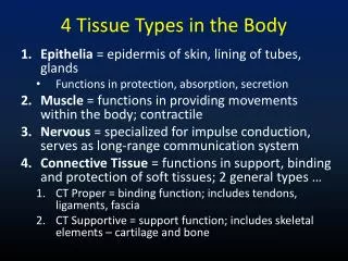
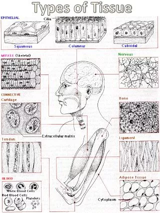
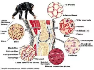
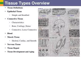
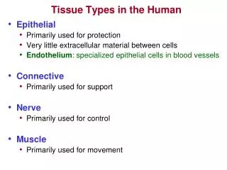
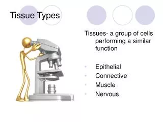
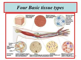
Tissue Types in the Human. Epithelial Primarily used for protection Very little extracellular material between cells Endothelium : specialized epithelial cells in blood vessels Connective Primarily used for support Nerve Primarily used for control Muscle Primarily used for movement.

E N D
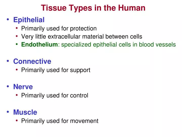
Tissue Types in the Human • Epithelial • Primarily used for protection • Very little extracellular material between cells • Endothelium: specialized epithelial cells in blood vessels • Connective • Primarily used for support • Nerve • Primarily used for control • Muscle • Primarily used for movement
Epithelial Tissue • Cells are polyhedral (many sided) with little interstitial space • Covers the outermost layer of tissue (skin) • Skin • Covers innermost and layer of most organs and cavities • Lungs, GI tract, Urinary tracts, Reproductive tracts • One side always exposed to: • Body exterior http://www.britannica.com/EBchecked/topic/190379/epithelium • Organ tract or cavity • Makes up the exocrine and endocrine glands • Exocrine (“excreting”): sweat glands, digestive glands, mammary glands • Endocrine (“hormones”): thyroid, pancreas, adrenal cortex • Cells have high regeneration potential but areavascular • Rely on perfusion for O2 supply • Many epithelial cells rest on a “Basement Membrane” • Basement Membrane = Basal Laminae + Reticular Laminae • Basal Laminae: flat “sheet” ofnonliving adhesive-like collagen and glycoprotein • secreted by the epithelial cells themselves • Reticular Laminae: “foundation” for the Basal Laminae
Simple Squamous Simple Cuboidal Simple Columnar Adjectives Describing Epithelial Tissue • Squamous (meaning “scale”) - flat cells • Cuboidal - cells as tall as they are wide • Columnar - tall and column shaped • Simple - having a single layer of cells • Stratified - having stacked layers • Transitional – “dome” surface cells • - capable of stretching (bladder) • Ciliated - cilia on the exposed surface • Examples you should remember: • SIMPLE SQUAMOUS EPITHELIUM • Permeable cell structure - used for filtration and gas exchange • Examples: capillaries, alveoli, kidney glomeruli • STRATIFIED SQUAMOUS EPITHELIUM • Used for “protection” • Examples: skin, inside of mouth, vagina • CILIATED COLUMNAR EPITHELIUM • Used to move substances along a particular direction using the cilia • Examples: upper respiratory tract, fallopian tubes Transitional Stratified Squamous Stratified Cuboidal Pseudostratified Columnar
Examples of Epithelial Tissue Simple Squamous Epithelium Artery Endothelium Stratified Squamous Epithelium Human Skin • Orange and brown/green covering: Adventicia • Blue: Actin in smooth muscle • Green: Elastic basal membrane (Basil Laminae) • Innermost Orange: Arterial Endothelium Ciliated Columnar Epithelium Tracheal Lung Tissue Cilia Ciliated Columnar Epithelial Cells
Diseases of Epithelial Tissue Simple Squamous Epithelium Arterial Endothelium Dysfunction The Beginnings of Atherosclerosis Tear in endothelial wall (injury - dysfunction) Monocyte (Macrophage) Cholesterol crystal deposits Red blood cell Foam cell (Lipid filled macrophages) Ciliated Columnar Epithelium Trachea Tissue from a SMOKER Fat deposits Note Lack of Cilia Note Disorganization of Columnar Epithelial Cells
ConnectiveTissue • Matrix - “non-living” component of connective tissue • Ground Substance • Proteoglycan aggregates (PGA) - pine tree shaped molecules • Glycosaminoglycans - neg charged binds Na+ & K+ attract H20 • Hyaluronic Acid - negative charged slippery polysaccharride • Condroitin sulfate • Fluid - H2O, gasses, nutrients for cells (H2O facilitates tissue “turgor”) • Minerals - Calcium salts • Adhesive glycoproteins – hold PGA’s together & to membranes • Chondronectin (cartilage), osteonectin (bone), fibronectin (fibrous tissue) • Laminin (holds epithelial cells to basement membrane) • Fibers • Collagen, Elastin, and Reticular Fibers http://www.siumed.edu/~dking2/intro/ct.htm • Cells - “living” component of connective tissue • “Blast” Cells, “Cyte” Cells, “Clast” Cells • Macrophages and white blood cells • Mast cells containing Heparin & Histamine • Adipose tissue
Skin Turgor The lack of turgor results in skin tenting
Proteoglycans Electron Micrograph of actual Proteoglycan Aggregate Other Glycosaminoglycans Dermatin sulfate Keratin Sulfate
Types of Fibers Collagen • Fibrous protein in connective tissue structure • Derived from Greek word meaning “to glue together” • Constitutes about 50% of the proteins in man • Present to some degree in all human organs • Collagen has a finite life span after which it is degraded to the constituent amino acids and replaced by new fibers. • Has high tensile strength: • 4.5 pound load needed to break collagen fiber 1 mm thick • Maximal strength of “scar” collagen is about 75% of the original tissue http://www.ncbi.nlm.nih.gov/books/bv.fcgi?rid=mcb.section.6542
Collagen Fiber (Fibril) Each collagen molecule (also called a tropocollagen) is connected to others viaPYRIDINIUM CROSS-LINK BONDS. These bonds are degraded in Ehlers-Danlos Syndrome, OsteogenesisImperfecta, and metastatic bone cancer and whose residue can serve as biomarkers which will be elevated in the urine. Microfibril A Collagen Molecule (Tropocollagen) Collagen Structure Each chain connected to the other two withHydrogen Bonds Collagen Molecule (Tropocollagen) Alpha Helix Chains Within Each Collagen Molecule Fibril Although Hydrogen bonds are weak, the “stacked intertwined” formation of the triple helix give collagen remarkable strength. Microfibril Fiber Tropocollagen Helix Individual Amino Acid Bonds Are Reinforced With Hydrogen Bonds
Diseases that Affect Collagen • Overproduction of Collagen Fibers • Lung Fibrosis (Cystic Fibrosis) – excess glandular secretions (mucous) • Caused by a mutation in CTFR gene product of this gene is ion channel • This channel is important in creating sweat, digestive juices, and mucous • High salt content in sweat is usually present in CF kids • Life-span used to be limited to 20-30 years….now possibly 40-50 years • Obstructions and fluid in lungs breathing disorders & numerous infections • Obstructions in pancreas digestive enzymes nutrient absorption • Malnutrition growth http://www.kidshealth.org/kid/health_problems/heart/cystic_fibrosis.html • Liver Cirrhosis – irreversible scarring (fiber deposition) in the liver • Common causes: Hepatitis-C Hepatitis-B, alcoholism • Alcohol blocks normal metabolism of protein, fats, and carbs injury • Cirrhosis edema & ascites (fluid in peritoneal space) • Liver cannot make Albumin blood looses osmotic (sucking) pressure • Cirrhosisinfection risk, jaundice, bruising & bleeding, portal hypertension • Cirrhosis will elevate Aminotransferase enzymes • ALT, AST, GGT(aka SGOT- large elevations associated with alcoholism) • Atherosclerotic heart disease http://www.digestive.niddk.nih.gov/ddiseases/pubs/cirrhosis/
Diseases that Affect Collagen • Autoimmune Disorders that Damage Collagen • Lupus Erythematosus-production of “auto antibodies” that target body tissue • 90% of Lupus patients will experience joint and muscle pain • Pain caused by collagen damage and destruction • Collagen damage and inflammation can occur anywhere in the body • Most common areas affected: skin (rashes that result from sunlight exposure), articular tissue, lungs, blood vessels, liver, kidneys, and nervous system • Course of the disease is unpredictable with attacks and remissions • Most common in women ages 18 – 40 http://www.merck.com/mmhe/sec05/ch068/ch068b.html • Insufficient Collagen • Ehlers-Danlos syndrome-rubber man - contortionist disease • Weakening of collagen cross-link bonds • OsteogenesisImperfecta- brittle bones • Scurvey-Vitmain C deficiency • Too few hydrogen bonds in the formation of the collagen molecule • Inferior tissue formation in bones, blood vessels, skin, and teeth
Elijah Price David Dunn
Therapy for Common Collagen Diseases • Cystic Fibrosis • Clearance techniques for excess lung secretions • Pancreatic enzyme replacement for pancreatic duct obstruction • Healthy diet and exercise • Drugs: • Ibuprofin – slows rate of decline of pulmonary function • Corticosteroids– inflammation in lungs, joints, and vasculature • Antibiotics – to treat and reduce the incidence of lung infections • Rotation of drugs to prevent development of resistance • Immunoglobulin experimental research (currently in human trials phase)
Therapy for Common Collagen Diseases • Lupus • Drugs: • Disease Modifying Anti Rheumatic Drugs (DMARDS): flare-ups • NSAID’s • Corticosteroids and other immunosuppresants • Hydroxychloroquine inflammation (used as an anti-malarial drug) • activation of inflammation causing dendritic cells (2009) • Experimental drugs • BENLYSTA (Belimumab)– antibody that inhibits B-lymphocyte stimulator (BLyS). BLyS turns B-lymphocytes into plasma cells which produce antibodies (first line of defense “foot soldiers” against infection). Overproduction of BLyS results in overproduction of antibodies. Excess BLyS production is noted in Lupus and other autoimmune diseases. (Phase 3 clinical trials in 2009) • Autograft bone marrow stem cell infusion very promising in reducing symptoms • Ultraviolet Radiation Therapy (Phase 2 clinical trials 2009)
Other Types of Fibers • Reticular Fibers: • Actually very fine collagen fibers • Usually form a network • Fill “space” between other tissues & organs • Contained in the reticular laminae • Elastic Fibers: • Contain protein called ELASTIN • Elastin molecules look like “coiled springs” • Return to original shape after distortion • Found in arteries, skin, alveoli of lungs (prominent in skin) • Synthesized by only fetal and juvenile fibroblasts • Loss of elastin with age contributes to development of “wrinkles” • Degraded elastin found in Aortic Aneurysm & COPD • Malformed elastin is found in Aortic Stenosis • genetic predisposition for this type of AS
Elastin Fibers • Stretched or taught • Relaxed
Types of Connective Tissue • Fibroconnective Tissue • Cartilage • Bone http://lifesci.rutgers.edu/~babiarz/dcttx.htm
Types of Connective Tissue • Fibro connective Tissue - matrix composed mostly fibers • Areolar-”Loose connective tissue” “packing material” holds organs in place • Most common connective tissue in all vertibrates • Component of some basal membranes (attaches underlying structures) • Separates muscles - allows for muscles to slide over each other • Fibers in extracellular matrix are collagen & elastin • Extracellular matrix is not well organized • Has more cells than Dense Regular or Dense Irregular • Reticular- form an internal skeleton for lymph, bone marrow, fat, & spleen • Fibers are collagen & elastin • Adipose- highly vascular insulator, shock absorber & energy store • Cells account for 90% of tissue mass (little matrix present) • Dense regular - closely packed parallel collagen fibers – few cells • Found where tension is exerted in a particular direction • Examples: tendons, ligaments • Dense irregular - closely packed non-directional collagen – few cells • Forms “sheets” where tension is exerted in many directions • Examples: dermis of skin, muscle fascia, organ & nerve coverings • Elastic- composed of mostly elastin fibers • Examples: vocal cords, ligamenta flava (vertebral connective tissue)
Fibrocyte Nucleus Collagen Fiber Elastic Fiber Areolar Tissue
Adipose Tissue X 200 ( bv = blood vessel ) ( arrow: adipocyte nucleus ) bv
Dense Regular Tissue (Tendons & Ligaments) Horse Tendon x100 arrow: orientation of collagen fibers
Dense Irregular Tissue (Dermis of Skin & Muscle Fascia)
Types of Connective Tissue • Cartilage - matrix mostly fibers & ground substance • Avascular (slow to heal) and not innervated • Composition of cartilage matrix: • Ground substance: chondroitin sulfate & hyaluronic acid • Collagen (the main fiber) & elastin • Perichondrium – surrounding tissue from which nutrients diffuse • Limits cartilage thickness - nutrients must diffuse entire tissue thickness • Gives rise to chondrocytes • Types of cartilage: • Hyaline - tough & flexible - much matrix / few cells - shock absorber • Covers ends of long bones (articular cartilage – eroded in OA) • Forms “skeleton” of trachea and bronchi • Fibrocartilage - less firm than hyaline - more cells and fibers • Similar in structure to dense regular tissue (tendon) • Transitional tissue between tendon and articular hyaline cartilage • Component of joint capsules & transitions to spongy knee menisci • Elastic - contains more elastin fibers than other types of cartilage • forms ear pinna & epiglottis http://education.vetmed.vt.edu/curriculum/vm8054/labs/Lab7/lab7.htm
Hyaline Cartilage X 250 arrows: Perichondrial borders Hyaline Cartilage Perichondrium on the left Chondrocytes form in the perichondrium and move out into the tissue
Elastic Cartilage (note numerous chondrocytes and elastic fibers)
Types of Connective Tissue • Bone- matrix mostly calcium and phosphate • 65% of bone weight is calcium hydroxyapatite • Calcium phosphate, calcium hydroxide, calcium carbonate • Tropocollagen subunits giving bone elasticity and fracture resistance • Bone collagen with age fracture risk • Highly vascular and well innervated • Contains lymph channels • Functions in mineral storage and blood cell production • Regulation of Ca++ metabolism - bone remodeling (deposition resorption) • bloodCa++ Parathyroidhormone(PTH)osteoclast activity • blood Ca++Calcitonin osteoblast activity • Red marrow: contains hematopoietic tissue - produces blood cells • Influences on Bone Growth: • Levels of Ca++, Phosphorous, Vitamin D, HGH, estrogen, testosterone • http://emedicine.medscape.com/article/1254517-overview
Review of Bone Histology Epiphyseal Plates Haversian System (osteon) Compact (Cortical) Bone Haversian Canal (contain blood vessels) Lamellae (concentric rings of hard bone) Marrow Osteocytes in Lacunae Endosteum Periosteum Volkmans Canal Canaliculi (connecting tunnels) Trabecular (Cancellous or spongy) Bone
Growth Plates (epiphyseal plate or the physis) in Long Bone • Example: young baseball pitchers throwing curve balls • Widening of proximal humeral physisischemia • Necrotic changes, asymmetric growth • Possible premature closing of physis http://bjsm.bmj.com/cgi/content/full/40/9/749?rss=1 http://www.medscape.com/viewarticle/408524_5
Growth Plate (epiphyseal plate) in Long Bone Hematopoietic Tissue zone of resting hyaline cartilage zone of proliferation zone of hypertrophy zone of calcification ossified bone Epiphysis (bone end) Diaphysis (bone shaft) Length Increase (Growth) Occurs Toward Diaphysis Chondrocytes Chondrocytes die upon calcification - blood vessels from diaphysis grow into the area Chondrocytes divide and stack on top of one another Red Bone Marrow
Bone Diseases & Treatments • Pagets Disease - enlarged deformed bones • bone resorption and deposition weak bones, bone pain, fractures, arthritis • Affects about 1% of adults - rarely diagnosed in people under 40 years of age • Symptoms: pain, pinched nerves tingling & numbness, leg bowing, hip & knee pain • Bones most often affected: spine, femur, pelvis, skull, clavicle, humerus. • Treated with Bisphosphonate drugs (see osteoporosis treatment) • Osteoporosis - bone density fracture predisposition • Osteopenia – bone loss not as severe as Osteoporosis • bone resorption in the presence of normal bone metabolism • both cortical (thick) and trabecular (porous) bone density, but affects cortical bone more • Women start losing bone density about age 40, men at age 60 • Over 28 million people in the U.S. have osteoporosis • 80% of this 28 million are women • Many women experience up to a 20% in bone mass by 5 to 7 years after menopause • 1 in 2 women and 1 in 4 men over age 50 will have an osteoporosis related fracture in their lifetime. • Common Fracture Sites: thoracic vertebra, • distal radius (close to wrist), femur neck (Hip Fracture) http://www.medicinenet.com/osteoporosis/article.htm
Bone Diseases & Treatments • Osteoporosis – continued) • Causes: • Prolonged treatment with corticosteroids • Anorexia nervosa • Inadequate diet, especially during pregnancy and breast feeding • Amenorrhea estrogen metabolism disturbances bone loss (Osteopenia) • Treatment: • Estrogen replacement (for postmenopausal women)?? • EVISTA (Raloxifene) - Selective estrogen receptor modulator that bone resorption • Calcium supplementation • Vitamin D supplementation • Bisphosphonate drugs (also called diphosphonates) • FOSAMAX, ACTONEL, BONIVA, AREDIA, RECLAST– osteoclast activity • Also used to treat bone cancer & other bone weakening diseases • CALCIMAR or MIACALCIN (Calcitonin) osteoblast activity • FORTEO (Teriparatide) a PTH analog: osteoblast activity • Intermittent exposure to PTH will osteoblast activity more that osteoclast activity • Weight bearing exercise can bone mineral density and falls • Kemmler et al. Arch Intern Med. 2010 Jan 25;170(2):179-85. • Osteomyelitis - bone inflammation & destruction • Caused by bacteria and fungi spreading from other infection sites • Symptoms: fever, localized warmth & swelling, localized pain • Treated with antibiotics
Goals Goal Achieved by How it works Primary Strategy ( - ies) 1. Reduce Torque Tape Decrease Damage 2. Reduce Pressure Tuli’s Less Pain 3. Increase Flexibility Stretch, Massage, splint Speed up Healing 4. Optimize Inflammation Ice, Aleve (NSAID) 5. Proper nutrition Protein, milk, vitamin/mineral supplement
Bone Diseases & Treatments • Osteoarthritis - Degenerative changes in cartilage & bone • Loss of articular cartilage (proteoglycan loss water loss compliance) in hands, hips, and knees • Roughening, pitting, & destruction in hyaline cartilage joint “stiffness” • IL1β & TNFαfrom chondrocytes collagen production + catabolism of articular cartilage • May result in the formation of osteophytes (bone spurs) or nodes (Bouchard’s nodes) • 80 – 90 % of people over age 65 have some evidence of osteoarthritis • Cause • Heredity (50% - 65%), infection, endocrine disorders, joint injury, overuse • Fracture or Ligament Injury bad joint alignment + instability “wear and tear” • Treatmenthttp://www.medicinenet.com/osteoarthritis/article.htm • Exercise - helps maintain ROM, healthy cartilage, strength and reduces pain • Rogind et.al. 1998; Gur et.al. 2002 • Immobilization can worsen the course of the disease • Weight loss for OA in weight bearing joints • NSAIDS and COX2 inhibitors for pain (COX 2 inhibitors ??? VIOXX CELEBREX) • Injections of HYALURONIN (hyaluronic acid) or new artificial injectable materials • Corticosteroid injections may be useful when inflammation is present • Joint replacement (when conservative therapy fails) • “Tissue engineering” to regenerate cartilage has had some success • Glucosamine & Chondroitin Supplements…. .definitive meta analysis study says no benefit • (Sawitzke et al, Arthritis and Rheumatism, 58: (10), 2008)
Osteoarthritis of the Knee Knee Arthroplasty Bone on bone in this resected tibial plateau Bouchard’s nodes Osteophytes
Bone Diseases & Treatments • Rheumatoid Arthritis (RA) – Autoimmune inflammatory disease • May be related to genetic factors, environment triggering autoimmune response, or infectious agents such as viruses, bacteria & fungi • Usually occurs between ages 25 & 55 and affects mostly young and • middle age females - may fluctuate substantially in severity • Rheumatoid factor (autoantibody) + globulins →immune complexes • Immune complexes activate the compliment system →inflammation • Involves synovial membranes of joints (most common manifestation) • Inflammation leads to swelling & thickening of synovial membrane (↑ESR) • Joints most often affected:wrists, fingers, knees, feet, and ankles • May possibly affect: http://www.arthritis.org/disease-center.php?disease_id=31 • Heart – endocarditis, pericarditis, CHF, valvular fibrosis, MI • - RA and other autoimmune disease patients have an ↑risk for CHD • Lungs – fibrosis and pleural effusion • Kidneys – amyloidosis (deposition of insoluble proteins in organ tissue) • GI tract – anemia resulting from chronic disease and constant NSAID use • - most RA patients are anemic • Fibrin deposition (fibrosis) and necrosis are also present • 60% of RA patients are unable to work 10 years after disease onset • Most research suggests that life span is reduced 5 – 10 years
Bone Diseases & Treatments Rheumatoid Arthritis Comparison of Normal & Rheumatoid Joint Normal Joint Rheumatoid Joint Synovial Membrane Infiltration of numerous cell types Pannus (granulation tissue) Leukocyte Macrophage Interdigitating Cell (“traps” antigens) Cartilage T-lymphocyte or B-lymphocyte Neutrophil Capillary Bud (angiogenesis) Immune Complex Inflamed Synovial Membrane
Bone Diseases & Treatments Rheumatoid Arthritis
Bone Diseases & Treatments • Treatments for Rheumatoid Arthritis • NSAID’s • COX2 inhibitors • Corticosteroids • Disease-Modifying Anti-Rheumatic Drugs (DMARD’s) • Methotrexate • ↓TNF, neutrophils, histamine, lymphocyte number & function • ↓growth of certain cells in blood, skin, GI tract, & immune system • Cytotoxic + inhibits metabolism →↓immune function • Developed in 1940s’ as chemotherapy for Leukemia • Sulfasalazine - ↓immune function • PLAQUENIL (Hydroxychloroquine) –an antimalarial drug • Gold salt injections • Exercise to maintain joint mobility • Physiotherapy, physical therapy, water exercise • Surgery: synovectomy or joint replacement • Gene therapy: injection of genes that produces desired protein via a vector(vector: various types of viruses)
New Anti-Arthritic Drugs – “Biological Agents” (or “Biologics”) • Tumor Necrosis Factor (TNFα) & Interleukin blockers: • Must be given by subcutaneous injection or IV TNFα HUMIRA adalimumab REMICADE infliximab ENBREL etanercept • Mechanism (Effects): • Binds to TNF • Prevents attachment to its receptor • Inhibits inflammatory mediators • ↓inflammation in joint →↓pain $15,000 - $45,000 / year (2008) • Indications: • Rheumatoid Arthritis • Effective in 70% of patients who • have not responded to Methotrexate • Ankylosing Spondylitis • Psoriasis • Psoriatic Arthritis- 12% of people with • psoriasis have psoriatic arthritis • Crohn’s Disease - autoimmune • inflammatory bowel disease http://www.webmd.com/rheumatoid-arthritis/guide/biologics • Adverse effects: • Immunosuppression !! • ↑risk of infection !! • Tuberculosis common • Allergic reactions • KINERET Anakinra • Mechanism: Injectable man-made protein that blocks interleukin-1 (IL-1) • IL-1 →cartilage degradation, ↑bone resorption • Adverse Reactions: Injection site reactions, systemic infections (↓immunity) • malignancies, neutropenia
Ankylosing spondylitis: Ankylosing (fusing) Spondyl- (spine) –itis (due to inflammation)
Blood & Body Fluids • 62.5% of total body fluid is intracellular (contained within cells) and is abbreviated ICF • 37.5% of total body fluid is extracellular (outside of cells: blood, interstitial fluid, etc) abbreviated ECF • Average human blood volume is about 5 Liters • 3 Liters plasma + 2 Liters RBC’s, WBC’s etc • Hematocrit (“crit”): packed RBC volume – about 45% of total volume for men, 40% for women • Anemia: crit < 40% for men or < 30% for women ( [Hb] < 14 g/dL for men, < 12 g/dL for women ) • Anemia can lead to fatigue & weakness and may be caused by: • Colon Cancer (bleeding), IBS (bleeding), Bleeding Ulcer, Crohn’sDisease (bleeding) • Other Cancers • ↑cytokines (TNFα, IL-1 →↓EPO synthesis →↓RBC count) • Chemotherapy & Radiation for cancer • RBC destruction + ↓RBC production • Kidney disease • ↓Erythropoietin (EPO) + Uremia →↓RBC’s and ↓platelet function • Blood Components • Formed elements: RBC’s, WBC’s, Platelets • Plasma: Plasma = Serum + clotting proteins such as fibrinogen (Plasma =Serum + Proteins) • Elevated fibrinogen levels →↑risk of stroke and atherosclerotic disease (tests available)
Blood & Body Fluids • Red blood cells (Erythrocytes) - formed in bone marrow • Production and homeostasis regulated by tissue oxygenation • ↓ tissue O2→↑erythropoietin from kidneys & liver →↑RBC’s • White blood cells (Leukocytes) • Primary effector against infection & tissue damage • WBC’s engulf foreign substances & lysosomalenzymes digest them • Inadequate lymphatic function or circulatory flow away from tissues →abscess • GRANULOCYTES (Polymorphonuclear cells) - granulated WBC’s • Neutrophils - 62% of WBC’s - 1st to travel & arrive at injury – “kamikazi” phagocytotic • Eosinophils - 2% of WBC’s - destroy parasites - involved in allergies • Basophils - < 1% of WBC - release histamine & heparin • AGRANULOCYTES – phagocytotic non-granulated WBC’S • Monocytes - 5.3% of WBC’s - become lysosome filled macrophages • Play important role in remove dust and necrotic tissue in lungs • Macrophages - monocytes that have left the circulation – long lived • Macrophages release cytokines and compliment proteins (inflam. mediators) • Lymphocytes – T cells & B cells – 15%-40% of WBC’s - function in acquired immunity • Antigen (pathogen) →B cell activation →antibody production • Helper T cells →cytokines →direct or mediate immune function • Cytotoxic T cells → cytotoxic granules →death of pathogen infected cells http://thyroid.about.com/library/immune/blimm06.htm
Macrophage: (in the box) Arrow: Pedicle for locomotion: Monocytes
large arrow: Basophil small arrow: Neutrophil Arrow: Eisonophil