HEPATITIS
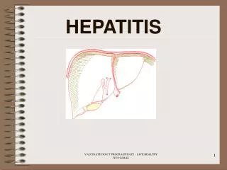
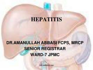
HEPATITIS. DR.AMANULLAH ABBASI FCPS, MRCP SENIOR REGISTRAR WARD-7 JPMC. The liver is designed to maintain body’s chemical and metabolic homeostasis. THE LIVER. - The liver is the largest organ in the body, occupying The entire upper right quadrant of the abdomen.

HEPATITIS
E N D
Presentation Transcript
HEPATITIS DR.AMANULLAH ABBASI FCPS, MRCP SENIOR REGISTRAR WARD-7 JPMC DR.AMANULLAH
The liver is designed to maintain body’s chemical and metabolic homeostasis DR.AMANULLAH
THE LIVER -The liver is the largest organ in the body, occupying The entire upper right quadrant of the abdomen. - Performs over 500 vital function. - It processes all nutrients the body requires - The liver manufactures bile, - One of the liver's major contribution is to criminate the toxic substance Old red blood cells are removed from the blood by the liver and spleen - Damage to the liver can impair these and many other functions. DR.AMANULLAH
THE LIVER LINED WITH TWO TYPES OF CELLS : Acini cell :it is the functional unit of liver . Kupffer's cell, monocytes, lymphocytes: -It is apart of reticular endothelial system [ monocytes, macrophage], its phagocytic cells that are capable of removing old and defective blood cells , bacteria, and other foreign material from portal blood. -This phagocyte action remove the enteric bacilli and other harmful substances that filter into the blood from the intestine . DR.AMANULLAH
Parenchymal Microanatomy DR.AMANULLAH
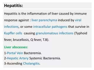
HEPATITIS Hepatitis is inflammation of the liver that can be caused by -Viruses ( e.g. Hepatitis B or hepatitis C ) , -Medications ( e.g. methyldopa or isoniazid ) , or -Immunologic abnormalities ( e.G., Autoimmune hepatitis) -Hepatitis can occur as acute or chronic disease. *Chronic disease generally is defined as that persisting for 6 months or longer . DR.AMANULLAH
Viral Hepatitis -Symptoms and time course alone cannot be used for differentiating between the virus types -Therefore, viral serologies are needed to distinguish between infections caused by each of the viruses. DR.AMANULLAH
HEPATITIS VIRUSES DR.AMANULLAH
HEPATITIS A -It is most common type of viral hepatitis occurring world-wide symptoms , of infection with HAV are usually acute in onset and infection occur primarily in children and young adult Character of virus: -In small RNA virus that can be detected in the liver , feces and blood during the late incubation periods and preicteric phase of illness . DR.AMANULLAH
EPIDEMIOLOGY -From the time of infection to the development of hepatitis symptoms such as jaundice, malaise and right upper quadrant pain. -The contagious period for hepatitis A appears to start 1 to 2 weeks) before onset symptoms and end strongly (10 to 15 days) thereafter. -Because high concentrations of hepatitis A are found in the stool of infected patients, usually mode of transmission is by directed fecal- oral contact or fecal contamination of food or water, there is no carrier state for hepatitis A. DR.AMANULLAH
SINGS AND SYMPTOMS Hepatitis A is associated with: -Fever (in 50% of patients) -Nausea -Anorexia -Malaise -Abdominal discomfort -Dark urine -Jaundice DR.AMANULLAH
PATHOPHYSIOLOGY -Hepatitis A infections are asymptomatic in approximately one third of patient, and other third develop mild nonspecific symptoms, and last develop more sever symptom including jaundice. -Patient with more sever symptom have an increase in theirserum aspartate aminotransferase (AST), alanine aminotransferase (ALT), and bilirubin, which resolve over 4 or 6 months. DR.AMANULLAH
DIAGNOSIS - Can not be made on clinical symptoms alone. - Diagnosis is based on measurement of immunoglobulin M ( IgM) antibodies to hepatitis A in the serum . -(IgG) antibodies to hepatitis A appear in the serum and against reinfection. DR.AMANULLAH
Serological test With the onset of jaundice, antibody to HAV (anti-HAV) become measurement in the serum. 1- Level of anti- HAV antibody of immunoglobulin M (IgM) Class risen sharply indicate acute infection. ( IgM : Onset of jaundice – short half life) 2- Then after acute illness , anti – HAV antibody of the immunoglobulin G (IgG) class predominate and remain indefinitely indicate past stage of HVA infection. ( IgG: last stage – long half life-chronic, recovery stage ) DR.AMANULLAH
Hepatitis B VIRUS DR.AMANULLAH
Hepatitis (B)(( serum hepatitis ))By DNA virus ETIOLOGY : HBV can be transmitted by: -Blood or sexual exposure. -Through the infected blood exposure contaminated fluid splashes in an eye or -When sterile techniques are not used with: intravenous needles.. Tattooing .. body piercing ... DR.AMANULLAH
ANTIGENS OF HBV -Three type of circulating antigen are present in the majority of patient : 1-Hepatitis B surface antigen .. HBsAg The outer coat of the hepatitis B virus 2-Hepatitis B core antigen .. HBcAg The core of hepatitis B virus that is produced and replicates in the hepatocyte nucleus<< cause tissue damage 3-HBeAgappears in the serum of the patient during the incubation period If the test are positive for hepatitis B e antigen the patient have high degree of infectivity. DR.AMANULLAH
SIGN AND SYMPTOM If it is acuteMalaise, Anorexia, Nausea, Vomiting, Jaundice Dark yellow urine ,Weakness Flu like symptom << before appearance of jaundice and dark yellow color If it is chronic fatigue , weakness and malaise is the sign. DR.AMANULLAH
PATHOPHYSIOLGY Incubation period : 17 -98 days -Acute hepatitisB associated with high elevation in AST and ALT Also in bilirubin -In chronic hepatitisis association with mild elevation in AST and ALT -With normal alkaline phosphatase and gamma glutamyl transpeptdase . -Platelet and RBC remain normal. DR.AMANULLAH
Diagnosis After infection with hepatitis B: -HBsAg can be measured in the serum 30 to 60 days after exposure to hepatitis B -So, antibodies against HBsAg developed and provide long-term immunity. -IgM antibodies against HBcAg develop and persist for about 6 months -So. HBcAg antibodies and IgM antibodies can use as markers for hepatitis B. -HBeAg is a marker of rapid viral replication. - -The develop HBeAg antibodies correlate with loss of replication virus and decrease infectivity of the virus. -Present of HBeAg, HBDNA indicate replication. DR.AMANULLAH
Hepatitis C By RNA virus ETIOLOGY: Infected blood exposures such as blood transfusions, needlesticks or tattooing . Also by sexual transmission and perinatal (mother or infant ) transmission. Incubation period :15 to 160 days PATHOPHYSIOLOGY : Serum ALT, AST and bilirubin increase but not high as in hepatitis B SIGN AND SYMPTOM :The same as hepatitis B DR.AMANULLAH
DIAGNOSIS -Hepatitis C can be diagnosis by detection of antibodies against the hepatitis virus or hepatitis C viral RNA in the patient Serum -It can be measured within 1week of infection. -Antibodies against hepatitis C can be measured 6 to 9 week after infection. -If the patient test positive for antibodies against hepatitis C and negative for hepatitis C viral RNA << mean the patient have resolved infection. DR.AMANULLAH
Differential features of viral hepatitiS DR.AMANULLAH
Hepatitis D ((delta)) -The virus is an incomplete or detective RNA .. nucleocapside core is thought to be coated with HBsAg - Hepatitis D infection can only occur as coinfection or superinfection with hepatitis B . Etiology : It can transmission by serum or I.V drugs users DR.AMANULLAH
DIAGNOSIS -Anti-delta IgM is detected during acute infection -HDAg and anti-HDAg disappear from serum when HBsAg is cleared -HD need HB in replication, HBsAg -Incubation 1-2 month DR.AMANULLAH
Hepatitis E Is the waterborne pathogen. The virus is RNA virus Etiology: It can transmission by fecal-oral route or by contact with contaminated food or water. Incubation period:1 to 2 month Symptom : Jaundice Fatigue Loss of appetite Diarrhea Nausea Fever Abdominal pain Occurs for Young to middle age adults DR.AMANULLAH
Hepatitis F Little is known about the virus , suggesting the need for further research Hepatitis G (HGV) The virus is RNA Etiology: -Transmission by sexual , blood transfusion, injection drug user , or by hemodialysis patient , accidental needle stick exposure blood-born means DR.AMANULLAH
PATHOGENESIS -Liver appears normal in size and color but some time slightly edematous, enlarge and tender edged to palpation -The characteristic picture is a combination of hepatocyte necrosis and regeneration , a diffuse mononuclear cell in filtration , hyperplasia of kupffer cells , and varying degree of bile stasis . -The mononuclear cells are primarily lymphocytes , with some oesinophils noted . DR.AMANULLAH
CONT…. Lobular disarray is apparent , probably due to the presence of ballooned hepatic plates, which obliterate the sinusoids , smudging of hepatocyte cell membranes , and the presence of inflammatory cell. Large hepatocytes with a .. ground glass.. appearance of the cytoplasm due to the presence of HBsAg are characteristic in chronic hepatitis B but are not seen in acute hepatitis B DR.AMANULLAH
-Although the liver cell plates may be disrupted leading to focal disruption of the reticulin network , this framework is generally preserved , since the necrosis tend to be limited to single of adjacent group cell. -In severe cases of hepatitis , this framework is destroyed leading to the characteristic pattern of bridging necrosis and a poorer prognosis -In severe cases of hepatitis , this framework is generally preserved , since the necrosis tend to be limited to single or adjacent group cells. DR.AMANULLAH
INVESTIGATION 1-liver biochemistry: a) Elevation of aspirate amino transferase (AST) > 500 UI/L and alanine aminotransferase (ALT) b) Elevation of lactate dehydrogenises (LDH) c) Alkaline phosphates: is usually normal or mildly elevation <300UI/L d) Bilirubin (in blood): is expected to be elevated and too wear as disease regress. ) Urinary bilirubin: persist through out the disease. DR.AMANULLAH
CONT… 2-Hematological test: Prothrombin time (PT): is prolonged in sever cases Erythrocyte sedimentation rate is raised due to cytokine. 3-Viral markers test (anti-body to HAV) Present of anti-HAV IgM indicate an acute infection. 4- Differential diagnosis: Clinically it is from all other causes of jaundice but in particular from after type of viral hepatitis DR.AMANULLAH
CLINICAL FEATURES Varies from fulminant hepatic failure to sub clinical from off to begin asymptomatic.. 1) Prodromal phase: – occur in all patients and may be present 1or 2 weeks before one set of jaundice (not all patient develop jaundice) - main feature at this time are malaise , lassitude , anorexia , headache, low-grad fever , and for smokers the less of desire to smoke . patient may have discomfort in the right upper guardant . - Many patient experience arthrolgia , arthritis ,urticaria ,and transient skin rashes .. DR.AMANULLAH
Clinical featurecont… 2) Icteric phase: The onset of jaundice usually lasts 4 – 6 week in addition to that urine become dark and stool pale where the fever subside and appetite normalize and feel well , the liver become moderately enlarge and tender in addition may have spleenomegaly or tender lymphadenopathy . 3) Recovery phase: If uncomplicated recovery begins 1 -2 weeks from jaundice and last 2 – 6 weeks. Easy fatigue ability is common .stool color normalize, jaundice lessens. Urine color lightens, speenomegaly take several weeks , lab take 3-6 months. DR.AMANULLAH
COMPLICATIONS * A few patient show rapid clinical deterioration following the acute jaundice due to fulminant hepatitis and massive liver necrosis. It is frequently associated with acute renal failure The most common complication of viral hepatitis is called chronic persistent hepatitis . * following acute viral hepatitis a few patient may develop chronic active or aggressive hepatitis in which there is piecemeal destruction of liver and development of cirrhosis DR.AMANULLAH
Thank you DR.AMANULLAH
Cirrhosis DR.AMANULLAH
Definition: Cirrhosis is characterized by a diffuse increase in the fibrous connective tissue of the liver, with areas of necroses and regeneration of parenchymal cells, imparting a nodular texture histologically. In its later stages, cirrhosis leads to such deformity of the liver that it interferes with hepatobiliary function and the circulation of blood to and from the liver DR.AMANULLAH
Cirrhosis Pathophysiology: Primary event is injury to hepatocellular- elements -Initiates inflammatory response with cytokine release->toxic substances -Destruction of hepatocytes, bile duct cells, vascular endothelial cells -Repair thru cellular proliferation an regeneration Formation of fibrous scar- Primary cell responsible for fibrosis is- stellate cell Become activated in response to injury and - lead to ed expression of fibril-forming collagen -Above process is influenced by Kupffer cells which activate stellate cells by eliciting production of cytokines -Initially, fibrosis may be reversible if inciting events are removed -With sustained injury, process of fibrosis becomes irreversible and leads to cirrhosis DR.AMANULLAH
Forms of cirrhosesEtiology, pathology, pathogenesis A-LAENNEC'S CIRRHOSIS: -The 1st change in the liver caused by alcohol is the gradual accumulation of fat within the liver cells (fatty infiltration). -The accumulation of fat reflects a number of metabolic disturbances, including excess formation of triglycerides, their decreased utilization in the formation of lipoproteins, and decreased oxidation of fatty acids. -The primary cause of liver damage is believed to the direct effect of alcohol on the liver cell, which is increased by malnutrition -Grossly the liver is enlarged, fragile, and greasy in the appearance and may be functionally deficient because of the large accumulation of fat DR.AMANULLAH
-In far-advanced cases of Laennec's cirrhosis, thick fibrous band formed at the periphery of many lobules, partitioning the parenchyma into fine nodules. -In the final stages the liver is shrunken, hard, and almost devoid of normal parenchyma, resulting in portal hypertension and hepatic failure. B-POSTNECROTIC CIRRHOSIS: Postnectotic cirrhosis presumably follow patchy necrosis of liver tissue, resulting in large and small degenerative nodules surrounded and partitioned by scar and interspersed with normal liver parenchyma. -About 75% of cases tend to progress and result in death within 1-5 years. DR.AMANULLAH
-It is account for about 25% of the cases have a prior history of viral hepatitis -Many patients have positive test results for HBAg, indicating that chronic active hepatitis may be an essential event. -A small percentage of cases stem from documented intoxication with industrial chemicals, poisons, or drugs(phosphorus, chloroform, carbon tetrachloride) -A peculiar feature of postnecrotic cirrhosis is that it appears to predispose the patient to the occurrence of a primary hepatic neoplasm of liver (hepatoma). C- BILIARY CIRRHOSIS Liver cell destruction that begins around the bile ducts gives rise to a pattern of cirrhosis known as biliary cirrhosis. -The most common cause of biliary cirrhosis is posthepatic biliary obstruction. DR.AMANULLAH
-Stasis of bile causes its accumulation within the liver substance with destruction of liver cells. - Fibrous bands begin forming around the periphery of the lobule. -The liver is enlarged, firm, finely granular, and has a green hue. -Jaundice is always an early and primary part of the syndrome, as are pruritis, malabsorption, and steatorrhea. -The cause of this condition associated with lesions of the intrahepatic bile ductules is unknown -bile capillaries and ductules contain bile plugs, and the liver cells frequently contain a green pigment. The extrahepatic biliary tract is not involved.- Portal hypertension as a complication is rare.- DR.AMANULLAH
Clinical manifestations cirrhosis is insidious in its development Early symptoms: -are vague and nonspecific and include lassitude, anorexia, dyspepsia, flatulence, a change in bowel habits (either constipation or diarrhea), and slight weight loss. Nausea and vomiting, especially in the morning, are common. -In most cases the liver is hard and palpable regardless of whether it is enlarged or atrophied. The major and late manifestation Develops as a result of two types of disorderedphysiology: DR.AMANULLAH
A-Manifestation of hepatocellular failure: -jaundice occurs when there is active inflammation of the liver bile ductules (cholangitis) -Endocrine disturbances . - spider angiomas, testicular atrophy, gynecomastia, pectoral and axillary's alopecia , palmar erythema are all considered to be caused by an excess of circulating estrogen -increased pigmentation of the skin is result from excessive activity of melanin stimulating hormone. -hematological disorder -(Nose bleeding, gingival bleeding, menstrual bleeding) result of decreased hepatic production of the clotting factor -anemia, leucopenia, thrombocytopenia are result from hyper slenism DR.AMANULLAH
-folate, vitamin b12 , iron deficiency secondary to blood loss, increase hemolysis of RBCs , patient more susceptible to infection -peripheral edema -hypoalbuminemia abnormal salt and water retention result from failure of the liver to inactivate aldosterone and ant diuretic hormone -sweetish odor May be result from inability to metabolize methionine -encephalopathy Abnormalities in metabolism of ammonia DR.AMANULLAH
B-PORTAL HYPERTENSION MANIFESTATION : -Portal hypertension is defined as a sustained elevation of pressure in the portal vein above the normal level. The primary mechanism for inducing portal hypertension, regardless of the disease, is increased resistance to blood flow through the liver. Increase in splanchnic arterial flow.- -decreased outflow through the hepatic vein and increase inflow combine to overload the portal circuit. -The back pressure in the portal system causes splenomegaly and is partly responsible for the accumulation of ascites. DR.AMANULLAH