Hepatitis:
Hepatitis:. Hepatitis is the inflammation of liver caused by immune response against : liver parenchyma induced by viral infections , or some intracellular pathogens that survive in Kupffer cells causing granulomatous infections (Typhoid fever, brucellosis, Q fever, T.B).

Hepatitis:
E N D
Presentation Transcript
Hepatitis: Hepatitis is the inflammation of liver caused by immune response against : liver parenchyma induced by viral infections, or some intracellular pathogens that survive in Kupffer cells causing granulomatous infections (Typhoid fever, brucellosis, Q fever, T.B). Liver abscesses: 1-Portal Vein Bacteremia. 2-Hepatic Artery Systemic Bacteremia. 3-Ascending Cholangitis.
N Viral causative agents for hepatitis are: 1- Professional-hepatitis viruses: Hepatitis A,B,C,D, and E; strong tropism to hepatocyte. 2-Non-Professional viruses that cause extra-hepatic diseases: Yellow fever viruses, E.B virus secondary to I.M, CMV, adenovirus, Herpes simplex, and VZV.
Hepatitis B virus: Pathogenesis: -Infection of hepatocytes. -T-cell mediated cytotoxicity: interaction between hepatocyte-MHC classI-HBcAg or HBeAgfragments and CD8+-TCR. -Kupffer cell response; release of cytokines and inflammatory mediators; chemotaxis. -Enhanced natural killer cell activity; cytotoxicity. -Interferon-α production; Up-regulates MHC-I expression and inhibits viral replication cycle. -HBsAg/Anti-HBsAg Antibodies complexes activate complement system damage.
Clinical presentation of Hepatitis B infection: -Incubation period. -Acute infection period: A-Pre-Ictericphase: (days to week): mild fever, anorexia, myalgia, and nausea. B-Icteric acute phase: (one to two months): Jaundice(yellowish coloration of mucous membrane, conjunctivae, and skin), enlarged and tender liver. C-Fulminanthepatitis: (In 1-2% of patients): Much more extensive necrosis of liver during icteric phase; elevated fever, abdominal pain, renal dysfunction, Lethal in 8% of cases.
Acute Hepatitis B infection n Virulent strain of hepatitis, Co-infection (HDV), Uncontrolled immunity and cytokines Limited cell-mediated and humoral immunity Effective cell-mediated and humoral immunity Chronic stage; Asymptomatic carrier N Chronic active hepatitis Resolution Minimal chronic hepatitis (persistent) Fulminant Hepatitis Liver Cirrhosis Hepatocellular Carcinoma
Diagnosis of Hepatitis B infection: -In incubation period and pre-icteric phase: The first indicator is HBsAg and HBeAg(envelope). -In acute icteric phase: Elevated Bilirubin(T,D), Liver enzymes (Transaminases), Bilirubinuria , elevated Anti-HBc Antibodies in serum. -In Convalescence phase: Anti-HBs Antibodies starts elevation in serum(positive). -In Chronic period: HBs Ag: positive Anti-HBc Antibodies: positive Anti-HBs Antibodies: negative.
Treatment and Prevention: -Reduction or elimination of HBV replication indicators could be performed by Interferon-alpha therapy. -Antiviral-nucleoside analogs that given orally have similar effect on viral replication (Lamivudine oradefovir: inhibition of viral reverse transcriptase). Prevention: 1-Active vaccine:HBsAg (children vaccination or others). 2-Passive vaccine: Serum Anti-HBsAg (exposed persons; needlestick , infants born to seropositive mother, sexual)
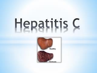
Hepatitis C virus infection: HCV accounted for ninety percent of cases of non-A, non-B hepatitis. Transmission: 1-It was initially identified as a major cause of post- transfusion hepatitis. 2-Intravenous drug users 3-Patients on hemodialysis. 4-Sexual route, and vertical. Pathogenesis: -Replication of virus in hepatocytes, lymphocytes, and macrophages. -Destruction of cells by viral replication cycle and immunity.
Acute hepatitis C infection Clinical outcomes of acute hepatitis C infection: Subclinical infection in 75% of cases Acute hepatitis C (25% of cases) Resolution of disease (months) Chronic hepatitis C (10-15 years) N Mixed Cryoglobulinemia Cirrhosis (20%) Hepatocellular Carcinoma Liver failure
N Diagnosis of HCV: -Detection of Anti-HCV-Recombinant viral protein antibodies in patient serum by ELISA. -Detection of viral nucleic acid in serum by RT-PCR technique. Treatment and Prevention: -Combination therapy of interferon-αand ribavirin provides a significantly improved response ( 30-50% for genotype 1 and 70-75% for viral genotype 2 and 3). -No available vaccine.
Hepatitis A infection: -HAV is responsible for most cases of infectious hepatitis. -Non-enveloped SS-RNA Virus. -Transmission: Fecal-Oral route or contaminated water. -Pathogenesis: -Absorbed into bloodstream by intestine; portal system of liver. -Replication in hepatocytes; destruction, exported into bile ducts; excreted into stool ( a high titer=1011viron/ml) -Released in bloodstream (to a lesser extent); transient viremia. -Acute hepatitis; inactivated vaccine (cell-cultured). Post-exposure prophylaxis (Anti-HAV-Abs).
Other causative agents for liver infection: Bacterial infections: Liver abscesses formation. 1-Portal-vein bacteremia. 2-Hepatic artery bacteremia. 3-Ascending cholangitis; E.coliis the most frequent agent 40% Facultative anaerobic and obligate anaerobic ascends from the duodenum. Several Parasitic infections: 1-Leishmaniasis: Leishmania donovani ; Visceral leishmaniasis (Kala-azar); jaundice, A/G ratio. In liver: intracellular Amastigotestage.
N 2-Extra-intestinal Amebiasis: Entamoebahistolytica Causes hepatic cyst due to transfer of trophozoite via portal system. 3-Schistosomiasis: Schistosoma mansoni; Schistosomula migrates to venous system of liver then to venous plexuses of large intestine. In liver: Egg-Granulomas in portal area. Pipe stem fibrosis of portal tracts. 4-Malarialife cycle: Plasmodium causes liver Schizonts.