Materials and methods
Evaluation of residual HIV-1 replication among individuals receiving different antiretroviral treatment regimens Giron , LB; Tenore , S; Gabriel, R; Janini , LMR; Sucupira , MCA; Diaz, RS. Department of Infectious Diseases, Federal University of Sao Paulo, São Paulo, Brazil. p=0,009* .

Materials and methods
E N D
Presentation Transcript
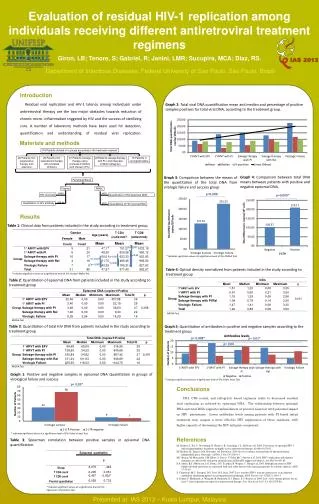
Evaluation of residual HIV-1 replication among individuals receiving different antiretroviral treatment regimens Giron, LB; Tenore, S; Gabriel, R; Janini, LMR; Sucupira, MCA; Diaz, RS. Department of Infectious Diseases, Federal University of Sao Paulo, São Paulo, Brazil p=0,009* p= 0,01* p=0,088 p= 0,008* p= 0,06 p= 0,09* Introduction Residual viral replication and HIV-1 latency among individuals under antiretroviral therapy are the two major obstacles towards reduction of chronic micro- inflammation triggered by HIV and the success of sterilizing cure. A number of laboratory methods have been used for detection, quantification and understanding of residual viral replication. Graph 2: Total viral DNA quantification mean and median and percentage of positive samples positives for total viral DNA, according to the treatment group. Materials and methods 116 Patientsdivided in 5 groups according to the treatment received Graph 4: Comparison between total DNA means between patients with positive and negative episomal DNA. Graph 3: Comparison between the means of the quantitation of the total DNA from virologic failure and success group PeripheralBlood 26 Patientsfirst suppressive therapy with efavirenz 25 Patientsfirst suppressive therapy with protease inhibitors 27 Patientssalvage therapy using protease inhibitors with ritonavir (PI-r) 22Patients salvage therapy with PI-r and integrase inhibitor raltegravir 16 Patientsin virological failure Plasma PBMC bDNA qPCR HIV viral load Quantitationof HIV episomal DNA BED kit Quantitationof HIV antibody qPCR Quantitationof HIV proviral DNA Results Table 1: Clinical data from patients included in the study according to treatment group. *p<0,00 * Indicates significant values at a significance level of 5%; ANOVA Test. *p<0,00 *p<0,00 *p= 0,025 *p<0,00 *p= 0,007 Table 4: Optical density normalized from patients included in the study according to treatment group * Indicates significant values at a significance level of 5%; Kruskal- WallisTest Table 2: Quantitationof episomal DNA from patients included in the study according to treatment group Graph 5: Quantitation of antibodies in positive and negative samples according to the treatment group. ANOVA Test ANOVA Test Table 3: Quantitationof total HIV DNA from patients included in the study according to treatment group * Indicates significant values at a significance level of 5%; Fisher Exact Test. ANOVA Test Graph 1: Positive and negative samples in episomal DNA Quantitationin groups of virological failure and success. Conclusions CD4, CD8 counts, and raltegravir based regimens relate to decreased residual viral replication as inferred by epissomal DNA. The relationship between episomal DNA and total DNA suggests replenishment of proviral reservoir with potential impact on HIV persistence. Lower antibodies levels among patients with PI based initial treatment may suggest a more effective HIV suppression of these regimens, with higher capacity of decreasing the HIV antigenic component. * Indicates significant values at a significance level of 5%; Fisher’sExactTest References M. Sharkey, I. Teo, T. Greenough, N. Sharova, K. Luzuringa, J.L. Sullivan et al. 2000. Persistance of episomal HIV-1 infectionintermidiates in patients on highly active antiretroviral therapy. Nat Med 6:76-81. M. Sharkey, K. Triques, D.R. Kuritzkes, M. Stevenson. 2005. In vivo evidence for instability of episomal human immunodeficiency virus type 1 cDNA. J Virol79:5203-10. M.J. Buzon, M. Massanella, J.M Llibre, A. Esteve, V. Dahl, M.C. Puertas et al. 2010. HIV-1 replication and immune dynamics are affected by raltegravir intensification of HAART suppressed subjects. Nat Med 16:460-65. P.A. Anton, R.T. Mitsuyasu, S.G. Deeks, D.T. Scadden, B. Wagner, C. Hung et al. 2003. Multiple measures of HIV burden in blood and tissue are correlated with each other but not with clinical parameters in aviremic subjects. AIDS 17:53-63. S. Cimerman, M.C. Sucupira, D.S. Lewi, R.S. Diaz. 2007. Less sensitive HIV-1 enzyme imunoassay as an adjuvant method for monitoring patients receivingantiretroviral therapy. AIDS Patient Care STDS 21:100-15. S. Palmer, F. Maldarelli, A. Wiegand, B. Bernstein, G.J. Hanna, S.C. Brun et al. 2008. Low- level viremia persists for ate least 7 years in patients on supressiveantirretroviral therapy. Proc NatlAcadSci U S A 105:3879-84. Table 3: Spearman correlation between positive samples in episomal DNA quantification. Presentedat IAS 2013 – Kuala Lumpur, Malaysia * Indicates significant values at a significance level of 5%; Spearman’sCorrelationtest