Download
1 / 57
570 likes | 601 Views
Enhance your CPR skills with this comprehensive guide, covering the history, techniques, and tools required for successful cardiopulmonary resuscitation. Understand the importance of airway control, chest compressions, and the use of automated external defibrillators in saving lives.

E N D
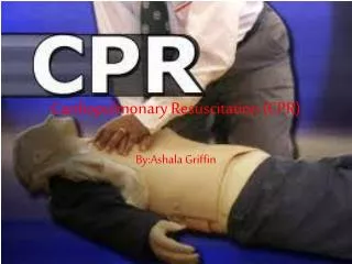
Cardiopulmonary Resuscitation ( CPR ) Houman Teymourian, M.D. Assistant professor, Department of Anesthesiology and Critical Care, Shahid Beheshti Medical University
GOAL • Artificial delivery of oxygenated blood to systemic circulatory bed at rates sufficient for preserving vital organ function and physiologic substrates until spontaneous circulation is reestablished.
History • Can be traced to at least Bibilical time • Contemporary approaches to CPR dates back to 1966 • 2000 Guide line: the first internationally recognized resuscitation guideline by AHA and European resuscitation council • 2007 Guide line: different aspects
BLS ( basic life support ) • Ventilation ( A-B) • Perfusion ( C ) • CPR Assessment • Automated External Defibrillation (D)
But first: • check responsiveness • check pulse • call for help and defibrillator
Assess circulation • 10 seconds • Carotid pulse in adults and children • Brachial in infants
Airway Control and Ventilation • Ventilation is critical for restoration of spontaneous circulation • Techniques used are dependent on the clinical situation
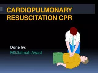
In out of hospital setting:- Mouth to mouth or mask to mouth- Head tilt, Chin lift, Jaw thrustThe major cause of upper airway obstruction in unconscious human is epiglottis move hyoid bone anteriorly
Open the Airway • Head-tilt/chin lift (no trauma victims) • The most common cause of obstruction is the epiglottis
Open the Airway • Jaw thrust (trauma)
Rescue Breathing : • 2 breath slowly ( 1.5 – 2 second ) after 30th compression during 1 or 2 person CPR until the airway is secured. • Slowly, to minimize high airway pressure and not to open esophagus. ( gastric inflation and aspiration )
Endo tracheal tube is the standard of airway control • Alternatives: - ( LMA ) laryngeal mask airway - ( C.T. ) Combitube - ( Ph.T ) Pharyngeal tube Not protecting from aspiration
Chest compression ( perfusion ) • Delivery of oxygenated blood during cardiac arrest • Recommended rate: 100/min • Depth: 1.5 to 2 inches ( 25 pounds ) • Must generate a palpable carotid or femoral pulse
Compression to relaxation: 1:1 • In 1 person CPR 15:2 compression - ventilation • In 2 person CPR 15:2 when airway is not secured • In 2 person CPR 5:1 when airway is secured • Continuous chest compression + asynchronous ventilation
Physiology • Direct cardiac compression is the main mechanism of blood flow ( cardiac pump mechanism ) • Increased intra thoracic pressure is another ( thoracic pump mechanism )
Other ways of chest compression • IAC ( Interposed Abdominal compression ) • Aortic diastolic pressure - Coronary blood flow • Vest CPR ( chest circumferential bladder ) • Active compression – de compression ( IRON Lung ) * Classic CPR is recommended.
CPR assessment ( monitoring performance of CPR) • Palpation of carotid or femoral pulse is not an objective evidence of the effectiveness of cardiac out put and only shows the transmission of a pressure wave in to the arterial tree. • Pupil size are of prognostic values constricted, or dilated but contracting to light are associated with a greater success in CPR and neurological out comes.
Direct systemic arterial pressure monitoring is gold standard ( Central arterial catheter ) • Aortic diastolic pressure = coronary perfusion pressure • P Et CO2 : is a precise of lung perfusion and cardiac out put.
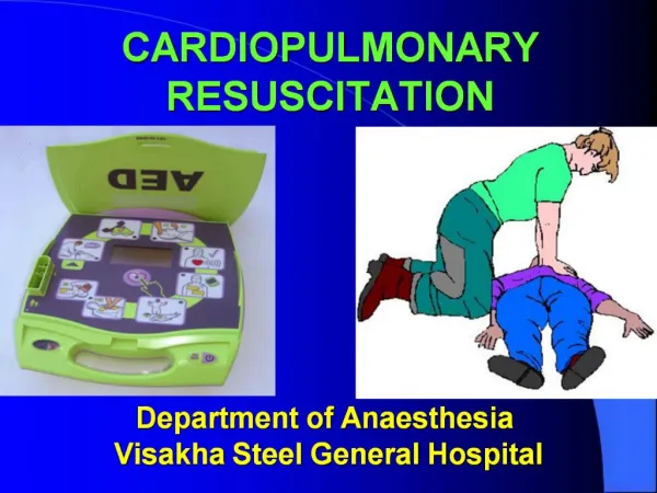
The automated external defibrillator • Most frequent cardiac rhythm in witnessed arrest in adult is VF. • CPR prolongs the duration of VF but cannot convert the arrhythmia to an organized rhythm. • AED is capable to recognize VF and VT
A E D • was first introduced in 1979 • AED is a part of BLS now • Chest compression must be stopped • 3 shocks is given automatically • 150 J Biphasic non escalating ( equivalent to monophasic 200 – 300 J ) • Must take place in less than 5 min in public places
ACLS( Advanced Cardiac Life Support ) • ACLS is a body knowledge and skill with which a physician must be thoroughly familiar • In a study using a computer the simulates cardiac arrest Only 30% of physician managed a simulated cardiac arrest in accordance with AHA ACLS guide line
Time since the last ACLS training is an important predictor of proper management of cardiac arrest. • Within 6 months 71% successful management • 6 month to 2 years 30% successful management • Longer than 2 years non
Management of cardiac arrest • Prompt recognition and treatment of potentially life threatening arrhythmia are essential components of ACLS • Has two compounds: electrical and pharmacologic
Pulse less ventricular tachycardia and ventricular fibrillation • Therapeutically are considered the same • The most common form of cardiac arrest • The greatest likely hood of short and long term survival • Definitive intervention is rapid defibrillation
Continue chest compression • Secure air way( ETT) • Hyper ventilate • Obtain IV access • but do not delay defibrillation
Defibrillation Two practical and important considerations are: • Energy out put Termination of VF is critically dependent on the amount of energy. 2. Resistance to current flow Flow is inversely related to resistance to minimize resistance: - one must use proper electrode position ( sternum and apex ) • Firm pressure ( 11 kg/each paddle ) • Optimal chest wall medium self adhesive paddles Electrode paste • End expiration defibrillations
Defibrillation • 150 J biphasic or 200 J monophasic followed by 200 – 300 J and third one of 360 J • 3 shock should be delivered rapidly • If witness but not monitored arrest is seen a single pericardial thump can be applied • If VF recurs the series of three shocks must be repeated • If VF persist drug therapy
So • Airway and ventilation ( ETT and 100% O2 ) • Defibrillation take precedence over intubation if defibrillator is ready • Drugs • chest compression continues
Routes of Drug Administration • Peripheral veins • Central veins • Tracheal • Intraosseous • Intracardiac
Peripheral Venous Route • Peak effect 1.5-3 min. after injection at antecubital fossa • IV push • 20 ml NS flush after drug injection • circulation time by 40% • Comparable to drug delivery through a central vein
Central Venous Route • Faster, higher peak concentration and more potent effect compared to peripheral injection • Should be used if it is already in situ • Inserting a central line is associated with problems of interrupting CPR, bleeding arterial puncture and air embolism
Tracheal Route • Second line route due to impaired absorption and unpredictable pharmacodynamics • Need 2-3 times the IV dose, diluted to at least 10 ml in 0.9% NS • Non-ionic drugs only: • adrenalin, atropine, lidocaine and naloxone
Drugs • Epinephrine ( through CV line ) • Regardless of under lying rhythm has beneficial effects ( cerebral and coronary flow ) • Recommended dose: 1 mg( 10 cc – 1: 10000 ) every 3 to 5 min as long as cardiac arrest persist • Can be injected to tracheal tube • Recommended dose : 2 – 2.5 times the Iv dose • Half life: 3 to 5 min • Large dose: 0.2 mg/kg was thought to be beneficial but to day is not accepted in CPR
2. Vasopressin • Clinical data shows endogenous plasma vasopressin level is significantly higher in patient successfully resuscitated versus in those who died • Acts on V1 smooth muscle receptor results in longer vasoconstriction and increase in coronary flow • Even in sever acidosis • It is an alternative to adrenaline • Recommended dose 40 unit/only one time • Has longer half life ( 10 – 20 min )
If VF Persists • Third shock is delivered ( 360 J mono or 200 J Biphasic ) Then if VF persists: Lidocaine 1.5 mg/kg or amiodarone 300 mg IV • Lidocaine enhances ventricular defibrillation ( lower energy, fewer shock ) but it is controversial AHA: it is an intermediate intervention without evidence of benefit or harm
Amiodarone • Amiodarone Prolongs the action potentials and refractoriness in cardiac tissue • Is superior to lidocaine to end VF but does not increase discharge rate • Rapid infusion of 300 mg in 20-30 ml NS IV push (cardiac arrest dose) • If VF/pulseless VT recurs, • Supplementary doses of 150 mg IV by rapid infusion • Followed by 1 mg/min for 6 hours and then 0.5 mg/min • Maximum daily dose of 2 g
No response? • Consider: • Procainamide 100 mg every 5 min up to 17 mg/kg (750-1500 mg) • Magnesium sulfate 1 gr IV every 5 min up to 4 gr • Phenitoin 50 mg/min Iv (250 mg) • Esmolol 0.3 mg/kg/min Iv infusion • Isoproterenol
If VF persists: Sodium bicarbonate infusion if 1. Pre existing acidosis 2. Acidosis documented with ABG 3. Hyperkalemia
Sodium bicarbonate • no benefit in survival of VF • adverse effect are documented: Plasma hyperosmolality Paradoxical cerebrospinal fluid acidosis CO2 generation vasodilatation hypotension In cardiac arrest pulmonary blood flow Co2 transport and elimination =Metabolic + respiratory acidosis * Bicarbonate should not be used routinely in treatment of cardiac arrest
It is used if prolonged VF arrest exist ( at least 15 min ) • Initial does: 1 mEq/kg • Followed by: 0.5 mEq/kg every 10 min • ABG- treat can be based on measurement
( PEA ) Pulseless Electrical activity • Refers to heterogeneous group of cardiac rhythm characterized by pulseless ness in presence of some type electrical activities other than VF or VT • Included electromechanical dissociation ( Idioventricular rhythm, Bradyasystole) 3. High priority must be given to identification of a correctible cause.
H’s Hypovolemia Hypoxia Hydrogen ion (acidosis) Hyperkalemia/ hypokalemia/ metabolic disorders Hypothermia/ hyperthermia T’s Toxins/tablets (drug overdose) Tamponade, cardiac Tension pneumothorax Thrombosis, coronary Thrombosis, pulmonary Possible Underlying Reversible Causes
In any form of PEA A -B- C inject Epinephrine ( and atropine if bradycardia is present) • Pacing may be indicated • Find the cause