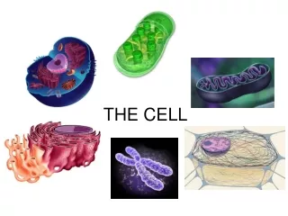
The Cell
The Cell. By Alex Vo and Adnan Abbuthalha. What’s a cell?. All living things are broken down into tiny little dots called “cells”. These dots are so small you can’t even see them. Yet, they are the what you need to survive on Earth.

The Cell
E N D
Presentation Transcript
The Cell By Alex Vo and Adnan Abbuthalha
What’s a cell? • All living things are broken down into tiny little dots called “cells”. These dots are so small you can’t even see them. • Yet, they are the what you need to survive on Earth. • Cells hold all of your biological needs in order to keep you a living thing. • If you don’t have them, then you might as well disappear into thin air.
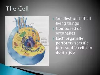
What does it do? • The purpose of cell is to create life and maintain it. • Each cell has a different function that helps our body develop healthily. • These tiny things make up our whole entire body from scratch and makes us into a walking, running thinking machine (not really).
Bacteria Cells make up the bread and mushrooms you eat The Diversity of Cells Cells come in all sorts and sizes. They perform many types of jobs in order to keep YOU alive. Muscle Cell produces energy so that you can run and have fun Red Blood Cell carry blood through your body White Blood Cell fight bad bacteria that can get you sick.
Organization of a Cell • Most cells are classified as a prokaryotic cell or a eukaryotic cell.
Prokaryotic Cells and Eukaryotic Cells • A prokaryotic cell is different from a eukaryotic cell because of the fact that it has no nucleus. It’s like a dumb cell with no brain. But, besides the nucleus, everything else is the same as a eukaryotic cell..
Plant or Animal Cells? • For a prokaryotic cell, there’s always going to be a cell wall, yet for a eukaryotic cell, it’s different. • Eukaryotic cells are divided into plants and animal cells. The difference is the fact that plant cells have a cell wall and an animal cell does not. • And obviously plant cells make up plants and animal cells make animals and humans.
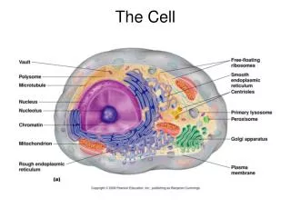
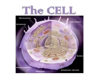
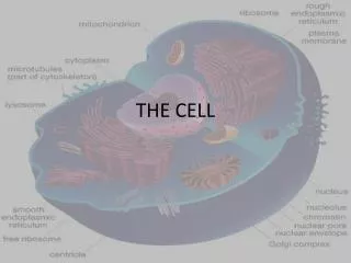
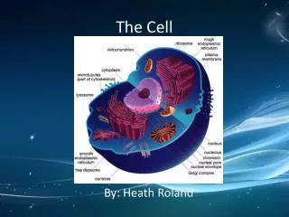
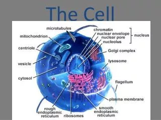
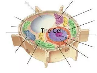
The Cell Structure Mitochondria Rough Endoplasmic Reticulum Lysosome Cell membrane Centriole Cytoskeleton Microtubule Ribosome Golgi Apparatus Microfilaments Smooth Endoplasmic Reticulum Please click on a button in the picture ONLY IN THE PICTURE OR ELSE YOU’LL MESS THINGS UP: Plastids Nucleus Nuclear Envelope Works Cited Cell Wall
What energizes cells? The Mitochondria • They are the batteries of a cell and keeps it constantly running. ( think energizer bunny) • 1-1000 mitochondria can exist in a cell depending on how hard its job is • It’s job is to gathers energy, kind of like your stomach, which takes in nutrients, breaks them down, and change them into energy that the cell can use (ATP). • The way this all occurs is Cellular Respiration.
How does cellular respiration work? • Cellular respiration is the way that food is broken down into ATP (energy) with oxygen.
How does cellular respiration work? That’s the glucose First, the glucose( sugar from the food that you ate) is brought through the outer membrane into the inner membrane
How does cellular respiration work? Next, it enters the matrix where it’s like a river full of water and enzyme( proteins that causes chemical reactions). From there, oxygen from the water is added and at the same time the enzymes are beginning to mix the glucose and oxygen together. This whole process is called Krebs Cycle( because it was found by some guy named Hans Krebs)
How does cellular respiration work? ATP Finally, it ends its journey at the end and exit the inner membrane and after that, the outer membrane. This time it’s not just any type of energy…. It’s ATP!
What Cleans up a cell? • A lysosome is another worker in the cell that has many enzymes which helps digest nutrients and other materials. • It can digest things like lipids, carbs, and proteins which mostly come from the food that you eat. • A lysosome also helps in removing debris and dead materials in a cell, kind of like a vacuum. • This all occurs with autophagy.
What’s autophagy Here are the steps: • First a membrane from the endoplasmic reticulum surrounds the unwanted material isolating it from the outside which prevents decomposing from spreading and infection. It’s like a gas chamber which keeps everything in. • Then, the lysosome comes in and attaches itself to the membrane. It forms a passage from itself to the organelle( big word for something in a cell). This passage is called an autophagic vacuole. • Finally the lysosome sends it’s deadly enzymes through the autophagic vacuole and destroys the organelle.
Cell Wall • A cell wall in only found in prokaryotic cells and eukaryotic plant cells • It’s made of strong fibers, made from carbohydrates (carbs.) and proteins, and woven together to be called cellulose • The purpose of a cell wall is to protect the cell from injuries and intruding materials that can harm the cell.
Ribosomes are found on the rough endoplasmic reticulum and are floating in the cytoplasm. Proteins are made here with special coded instructions from the nucleus, which makes all of them unique. Ribosomes
To make proteins, the two parts of a ribosome must connect with RNA (Ribonucleic Acid) from the nucleus. The RNA are lined with amino acids. These amino acids get stripped off with the instructions from the RNA. From there the proteins are created with the given info from the RNA. It’s basically like a protein factory and the RNAs are the messangers from the nucleus. How do ribosomes make protein?
The cell membrane is the thin, double layer of lipids( oils and fats from things that you ate) surrounding a cell. A cell membrane protects and helps support a cell while also allowing interaction with the outside. Cell/Plasma membrane
The cell membrane allows sodium, potassium, calcium, and a few other things to enter and exit the cell. One method of passing through the cell membrane is active transport. In this, particles move from areas of low concentration to areas of high concentration. Active transportation does require energy. Another way to put it is like moving from a less crowded room to a more crowded room. Passage through the Cell Membrane
Another method of transportation is called diffusion. In diffusion, molecules move from areas of high concentration to areas of low concentration. This makes sense since particles in general usually move to where there is more space. The diffusion of water is given the special term “osmosis”. It’s like moving from a crowded area to an area that isn’t densely populated. Diffusion and Osmosis
Vacuoles • It’s a single membrane surrounding the liquid or solid object • There’s nothing special about a vacuole, it’s just a sac that can hold many different types of materials. • There are many types of vacuoles in a cell. • In a plant cell, there’s a central vacuole that’s used to store water, which is important for the plant to survive. These are some cholesterol vacuoles. The central is the cholesterol. The green material surrounding it is the vacuole.
The Importance of a Central Vacuole in a Plant Cell • The central vacuoles stores salts, minerals, nutrients, proteins, and pigments (material that gives plants it’s certain colors). • All of these things in the vacuole are important especially the water, to the plant cell because it’s basically what keeps it from dying.
Centrioles • Cells do not give birth or reproduce, instead they divide in a way called mitosis. • Centrioles are things that help the cell to split up, kind of like forming a clone. • These cell parts are found in pairs and perpendicular, which means they form a 90 degrees angle. • They’re surrounded by groups of three microtubules. • Centrioles are mostly found in animal cells and not that often in plant cells.
What Do Centrioles Do in Mitosis? • First, the pair of cetrioles make a copy of itself, so there’s 4 now. • As the cells begin to split, each pair runs to the opposite ends of the cells. • Their microtubules shoot out and connect to each other forming a watermelon shape. • This formation will allow the chromosomes to divide into two groups evenly and be able to put them into orders once the cell splits.
Plastids • Plastids are only found in plant cells or other things that use photosynthesis (the way of gaining energy with the sun) • They’re found in the all around the cytoplasm( jelly like material surrounding a plant cell, after the cell wall). • A plastid’s job is to store molecules, which can vary depending on the type of plastid . • There are many types of plastids in a plant cell. • 3 types of plastids are chloroplast, chromoplast, and leucoplast.
Chloroplasts • Plants make their own food with these. • Chloroplasts are the areas where sunlight is gathered and turned into sugar for the plant to use. The process has to do with photosynthesis. • The stroma is the area where chemical reactions take place and sugar is created. • The thylakoid holds chlorophylls that gather the sun’s energy before being changed into sugar. • One thylakoid stack is called a granum. • The stroma lamellae is like the skeleton of chloroplasts that keeps it all together.
Photosynthesis in the Chloroplasts • First, the plant should’ve already absorbed water and carbon dioxide from the air around it. • Next,the chlorophylls in thylakoids absorb in the sun’s energy and sends that energy to the stroma. • The stroma begins the mixing of water and carbon dioxide. • Finally glucose ( sugar) is formed. • Oxygen is also created. That’s why people think it’s bad cutting down trees, because it’s lowering the amount of oxygen in the air. • It’s the opposite of how the mitochiondria works since the mitochondria breaks down food to create gluclose and chloroplasts build up to make glucose.
Chromoplast • It’s another plastid that holds a plants pigments (material that makes a plant’s color, mostly leaves). • It hold many pigments, yet does not hold any chlorophylls. • Colors are mostly red, yellow, and orange. • Color will vary depending on how much sun is able to be taken in. • The amount of sunlight depends on the seasons of the year, or how far the planted area is from the sun.
Leucoplast • Leucoplasts are non-pigmented plastids that are used in plants for storage There are three types: • Amyloplasts - colorless plant organelle related to starch production & storage • Aleuroplasts - colorless plant organelle related to protein production & storage • Elaioplasts - colorless plant organelle related to oil & lipid production & storage Amyloplasts Ealioplasts Aleuroplasts
The nucleus is pretty much the HQ of the cell. It controls cell processes by controlling the proteins made. The instructions for making every protein in the cell is found in DNA (Deoxyribonucleic Acid) RNA (Ribonucleic acid) is another nucleic acid that is connected with cell processes. The Nucleus
The nucleus envelope is the membrane that surrounds the nucleus. The small openings allow materials, like RNA and proteins, to pass through the nuclear membrane. The Nuclear Envelope
The Rough Endoplasmic Reticulum • It’s several connected membranes • The Rough ER makes proteins that are used in the cell membrane and also outside the cell membrane • It is called “rough” because of the ribosomes that are on its surface. These ribsomes send amino acid chains to the rough ER. • After the rough ER finishes making the proteins, it sends the proteins to the Golgi apparatus or cell membrane in vesicles, or tiny bubble transporters
It’s several connected membranes The smooth ER has a more tubular structure than the rough ER. The job of the smooth ER is to make and store lipids (ex.-steroids), and fatty acids. The Smooth Endoplasmic Reticulum
The Golgi apparatus is a stack of membranes that get proteins from the ER and change them. The Golgi Apparatus changes simple molecules into more complex ones and also alters proteins. It also assures that proteins don’t have flaws or unneeded materials. The Golgi Apparatus
Another task of the Golgi apparatus is to make lysosomes (small enzymye-filled organelles that break down carbohydrates, lipids, and proteins) In plants, the Golgi apparatus can make complex sugars. After the molecules inside the Golgi apparatus are ready for shipping, a vesicle is formed and sent out of the cell through the cell membrane. The Job of A Golgi Apparatus.
Some cells have a cytoskeleton that helps the cell keep its shape. The cytoskeleton also helps with cell movement. The two essential structures in a cytoskeleton are microfilaments and microtubules. Cytoskeleton
The cytoskeleton is connected to every part of the cell membrane and every organelle. Motor proteins that attach to organelles move them along microfilaments and microtubules almost like a train carrying cargo over tracks. More of the Cytoskeleton
Microfilaments are composed of a long, thin protein called actin. In muscle cells, actin and myosin (which are called actomysoin when together), help contract and relax the muscle cell (which in turn helps muscles to relax or contract) The pushing and pulling of microfilaments in the cell membrane help the cell move. Microfilaments
Microtubules are thick, round, proteins called tubulin. They are important in cell division by attaching to chromosomes and helping them split. Microtubules can combine to form flagella (which aid in cell movement) and cilia (which help single-celled organisms move around) They both help the cell move quickly in water. Microtubules
Works Cited • micro.magnet.fsu.edu/ cells/plants/cellwall.html • biology.unm.edu/.../ Summaries/Cell.html • micro.magnet.fsu.edu/.../ mitochondria.html • www.msad54.k12.me.us/.../ Energy/Mitochondria.htm • micro.magnet.fsu.edu/ cells/plantcell.html • www.umanitoba.ca/.../ lab3/biolab3_2.html • micro.magnet.fsu.edu/.../ animalmodel.html • www.alumni.ca/~mcgo4s0/ t1/cellsfinalfinal.html • www.Hybridmedicalanimation.com • http://www.ebi.ac.uk/microarray/biology_intro_files/cell.jpe • http://www.arvanitakis.com/en/bio/bacteria_structure.htm • http://www.emc.maricopa.edu/faculty/farabee/BIOBK/BioBooktransp.html • http://sun.menloschool.org/~cweaver/cells/c/cell_membrane/ • http://www.uga.edu/caur/nucleus.jpg • http://www.biologie.uni-hamburg.de/b-online/snowbird/snowbird/life_cell.htm • http://trc.ucdavis.edu/mjguinan/apc100/modules/TermsCells&Tissues/structures/images/ER.jpg • http://www.blc.arizona.edu/courses/181summer/graphics/graphics%20lect5/Life7e-Fig-04-11-0%20endoplasmic%20reticulum.jpg • http://schoolweb.missouri.edu/ashland.k12.mo.us/km/03page2/smooth.jpg • http://www.roche.com/pages/downloads/photosel/041123cd/Photo-Selection-Images/12.jpg • http://www.biologie.uni-hamburg.de/b-online/snowbird/snowbird/life_cell.htm • http://cwx.prenhall.com/horton/medialib/media_portfolio/text_images/FG01_15-02UN_90118.JPG • http://www.jdaross.mcmail.com/images/golgi3.jpg • http://folk.uio.no/laeide/pr_mitochondria.jpg • http://www.jdaross.mcmail.com/images/mitocho2.gif • http://www.jdaross.mcmail.com/images/lysosome.gif • http://www.cartage.org.lb/en/themes/Sciences/Zoology/AnimalPhysiology/Anatomy/AnimalCellStructure/Lysosomes/lysosome.jpg • http://www.stanfordphotonics.com/Life%20Sciences/LifeSci.htm • http://upload.wikimedia.org/wikipedia/commons/0/01/Rhoeo_Discolor_-_Plasmolysis.jpg • http://www.bms.ed.ac.uk/services/impact/pages/Image3.htm • http://focus.hms.harvard.edu/2001/May4_2001/Cytoskeleton.jpg • http://www.daviddarling.info/images/chloroplast.jpg
Work Cited • webnt.calhoun.edu/.../ Euk_and_Pro.htm • www.ou.edu/class/ pheidole/bacteria.html • www.esa.int/SPECIALS/ Eneide/SEMVJZRMD6E_1.html • www.mediscan.co.uk/ cfm/resultssearch.cfm?acti... • http://www.ebi.ac.uk/microarray/biology_intro_files/cell.jpe • http://www.arvanitakis.com/en/bio/bacteria_structure.htm • http://www.emc.maricopa.edu/faculty/farabee/BIOBK/BioBooktransp.html • http://sun.menloschool.org/~cweaver/cells/c/cell_membrane/ • http://www.uga.edu/caur/nucleus.jpg • http://www.biologie.uni-hamburg.de/b-online/snowbird/snowbird/life_cell.htm • http://trc.ucdavis.edu/mjguinan/apc100/modules/TermsCells&Tissues/structures/images/ER.jpg • http://www.blc.arizona.edu/courses/181summer/graphics/graphics%20lect5/Life7e-Fig-04-11-0%20endoplasmic%20reticulum.jpg • micro.magnet.fsu.edu/ cells/plants/vacuole.html • : fig.cox.miami.edu/.../ 150/cells/organelle.htm • www.lifesci.sussex.ac.uk/.../ vacuole.htm • nyecellshop.tripod.com/ id2.html • www.cartage.org.lb/.../ Centrioles/Centrioles.htm • www.mc.edu/campus/ users/rbuckley/carly.htm • www.mit.edu:8001/.../ other/esgbio/www/cb/org/ • library.thinkquest.org/ J0110902/Photosynthsis.htm • http://www.biochem.northwestern.edu/holmgren/Glossary/Definitions/Def-C/chromoplast.html • www.rsbs.anu.edu.au/.../ 10%2033%2001.htm • fig.cox.miami.edu/~cmallery/ 150/cells/plastid.htm • www.rsbs.anu.edu.au/.../ 10%2034%2001.htm • http://fig.cox.miami.edu/~cmallery/150/frames/thumbs/dblmb/plastids.content1.html • http://courses.washington.edu/dmandoli/Teams/CellWall/Research2000/Wcap100X.jpg • http://www.biologie.uni-hamburg.de/b-online/library/webb/BOT311/Cells_Tissues/pages/NuclearPoresLab.jpg • http://www.beyondbooks.com/bbx/tr/pv.asp?i=lif71/4d/00016722&u=http://science.uniserve.edu.au/mirror/biolproject/cell_bio/tutorials/pev/page3.html • http://www.beyondbooks.com/bbx/tr/pv.asp?i=lif71/4d/00017006&u=http://home.earthlink.net/~dayvdanls/lecw4cells7.html • http://www.microtubule.com/intracellular.jpg • http://www.imb-jena.de/~kboehm/mt-neg.jpg • http://www.tmd.ac.jp/artsci/biol/textbook/cytoskel.gif • http://www.beyondbooks.com/lif71 • http://science.howstuffworks.com/cell.htm • http://www.madsci.org/posts/archives/may2000/957278393.Cb.r.html • http://www.cellsalive.com
Works Cited • http://www.biology4kids.com/files/cell_main.html • http://www.biology-online.org/9/2_cell_structure.htm • http://www.cellsalive.com/cells/3dcell.htm • http://www.elmhurst.edu/~chm/vchembook/591cellstruct.html • http://www.phschool.com/science/biology_place/biocoach/cells/common.html • http://www.sirinet.net/~jgjohnso/cell.html • http://training.seer.cancer.gov/module_anatomy/unit2_1_cell_functions_1.html • http://www.trentu.ca/academic/biology/101/2.html • http://www.tvdsb.on.ca/westmin/science/sbi3a1/Cells/cells.htm • http://www.uky.edu/~rebeat1/cell.html • http://vilenski.org/science/safari/classifyall/eukaryotic.html • http://www.visionlearning.com/library/module_viewer.php?mid=64&l=&c3= • http://web.jjay.cuny.edu/~acarpi/NSC/13-cells.htm • micro.magnet.fsu.edu/. ../lysosomes.html • www.jdaross.mcmail.com/ lysosome.htm • :www.cartage.org.lb/.../ Ribosomes/Ribosomes.htm • library.thinkquest.org/ C004535/cell_wall.html