Download
1 / 76
890 likes | 1.59k Views
Circulatory System: The Heart. Circulatory System: The Heart. Gross anatomy of the heart Overview of cardiovascular system Cardiac conduction system and cardiac muscle Electrical and contractile activity of heart Blood flow, heart sounds, and cardiac cycle Cardiac output.

E N D
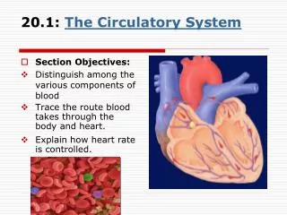
Circulatory System: The Heart
Circulatory System: The Heart • Gross anatomy of the heart • Overview of cardiovascular system • Cardiac conduction system and cardiac muscle • Electrical and contractile activity of heart • Blood flow, heart sounds, and cardiac cycle • Cardiac output
Circulatory System: The Heart • Circulatory system • heart, blood vessels and blood • Cardiovascular system • heart, arteries, veins and capillaries Two major divisions: • Pulmonary circuit - right side of heart • carries blood to lungs for gas exchange • Systemic circuit - left side of heart • supplies blood to all organs of the body
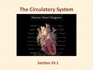
Position, Size, and Shape • Located in mediastinum, between lungs • Base - broad superior portion of heart • Apex - inferior end, tilts to the left, tapers to point • 3.5 in. wide at base, 5 in. from base to apex and 2.5 in. anterior to posterior; weighs 10 oz
Pericardium • Allows heart to beat without friction, room to expand and resists excessive expansion • Parietal pericardium • outer, tough, fibrous layer of CT • Pericardial cavity • filled with pericardial fluid • Visceral pericardium (a.k.a. epicardium of heart wall) • inner, thin, smooth, moist serous layer • covers heart surface
Pericardium and Heart Wall Pericardial cavity contains 5-30 ml of pericardial fluid
Heart Wall • Epicardium (a.k.a. visceral pericardium) • serous membrane covers heart • Myocardium • thick muscular layer • fibrous skeleton - network of collagenous and elastic fibers • provides structural support and attachment for cardiac muscle • electrical nonconductor, important in coordinating contractile activity • Endocardium - smooth inner lining
Heart Chambers • 4 chambers • right and left atria • two superior, posterior chambers • receive blood returning to heart • right and left ventricles • two inferior chambers • pump blood into arteries • Atrioventricular sulcus- separates atria, ventricles • Anterior and posterior sulci - grooves separate ventricles (next slide)
Heart Chambers - Internal • Interatrial septum • wall that separates atria • Pectinate muscles • internal ridges of myocardium in right atrium and both auricles • Interventricular septum • wall that separates ventricles • Trabeculae carneae • internal ridges in both ventricles
Heart Valves • Atrioventricular (AV) valves • right AV valve has 3 cusps (tricuspid valve) • left AV valve has 2 cusps (mitral, bicuspid valve) • chordae tendineae - cords connect AV valves to papillary muscles (on floor of ventricles) • Semilunar valves - control flow into great arteries • pulmonary: right ventricle into pulmonary trunk • aortic: from left ventricle into aorta
AV Valve Mechanics • Ventricles relax • pressuredrops • semilunar valves close • AV valves open • blood flows from atria to ventricles • Ventricles contract • AV valves close • pressurerises • semilunar valves open • blood flows into great vessels
Coronary Circulation • Left coronary artery (LCA) • anterior interventricular branch • supplies blood to interventricular septum and anterior walls of ventricles • circumflex branch • passes around left side of heart in coronary sulcus, supplies left atrium and posterior wall of left ventricle • Right coronary artery (RCA) • right marginal branch • supplies lateral R atrium and ventricle • posterior interventricular branch • supplies posterior walls of ventricles
Angina and Heart Attack • Angina pectoris • partial obstruction of coronary blood flow can cause chest pain • pain caused by ischemia, often activity dependent • Myocardial infarction • complete obstruction causes death of cardiac cells in affected area • pain or pressure in chest that often radiates down left arm
Venous Drainage of Heart • 20% drains directly into right atrium and ventricle via thebesian veins • 80% returns to right atrium via: • great cardiac vein • blood from anterior interventricular sulcus • middle cardiac vein • from posterior sulcus • left marginal vein • coronary sinus • collects blood and empties into right atrium
Nerve Supply to Heart • Sympathetic nerves from • upper thoracic spinal cord, through sympathetic chain to cardiac nerves • directly to ventricular myocardium • can raise heart rate to 230 bpm • Parasympathetic nerves • right vagal nerve to SA node • left vagal nerve to AV node • vagal tone – normally slows heart rate to 70 - 80 bpm
Cardiac Conduction System • Properties • myogenic - heartbeat originates within heart • autorhythmic – regular, spontaneous depolarization • Components • next slide
Cardiac Conduction System • SA node: pacemaker, initiates heartbeat, sets heart rate • fibrous skeleton insulates atria from ventricles • AV node: electrical gateway to ventricles • AV bundle: pathway for signals from AV node • Right and left bundle branches: divisions of AV bundle that enter interventricular septum • Purkinje fibers: upward from apex spread throughout ventricular myocardium
Structure of Cardiac Muscle • Short, branched cells, one central nucleus • Sarcoplasmic reticulum, large T-tubules • admit more Ca2+ from ECF • Intercalated discs join myocytes end to end • interdigitating folds - surface area • mechanical junctions tightly join myocytes • fascia adherens: actin anchored to plasma membrane; transmembrane proteins link cells • desmosomes • electrical junctions - gap junctions allow ions to flow
Metabolism of Cardiac Muscle • Aerobic respiration • Rich in myoglobin and glycogen • Large mitochondria • Organic fuels: fatty acids, glucose, ketones • Fatigue resistant
Cardiac Rhythm • Systole – ventricular contraction • Diastole - ventricular relaxation • Sinus rhythm • set by SA node at 60 – 100 bpm • adult at rest is 70 to 80 bpm (vagal inhibition) • Premature ventricular contraction (PVC) • caused by hypoxia, electrolyte imbalance, stimulants, stress, etc.
Cardiac Rhythm • Ectopic foci - region of spontaneous firing (not SA) • nodal rhythm - set by AV node, 40 to 50 bpm • intrinsic ventricular rhythm - 20 to 40 bpm • Arrhythmia - abnormal cardiac rhythm • heart block: failure of conduction system • bundle branch block • total heart block (damage to AV node)
Depolarization of SA Node • SA node - no stable resting membrane potential • Pacemaker potential • gradual depolarization from -60 mV, slow influx of Na+ • Action potential • occurs at threshold of -40 mV • depolarizing phase to 0 mV • fast Ca2+ channels open, (Ca2+ in) • repolarizing phase • K+ channels open, (K+ out) • at-60 mV K+ channels close, pacemaker potential starts over • Each depolarization creates one heartbeat • SA node at rest fires at 0.8 sec, about 75 bpm
Impulse Conduction to Myocardium • SA node signal travels at 1 m/sec through atria • AV node slows signal to 0.05 m/sec • thin myocytes with fewer gap junctions • delays signal 100 msec, allows ventricles to fill • AV bundle and purkinje fibers • speeds signal along at 4 m/sec to ventricles • Ventricular systole begins at apex, progresses up • spiral arrangement of myocytes twists ventricles slightly
Contraction of Myocardium • Myocytes have stable resting potential of -90 mV • Depolarization (very brief) • stimulus opens voltage regulated Na+ gates, (Na+ rushes in) membrane depolarizes rapidly • action potential peaks at +30 mV • Na+ gates close quickly • Plateau - 200 to 250 msec, sustains contraction • slow Ca2+ channels open, Ca2+ binds to fast Ca2+ channels on SR, releases Ca2+ into cytosol:contraction • Repolarization - Ca2+ channels close, K+ channels open, rapid K+ out returns to resting potential
Action Potential of Myocyte 1) Na+ gates open 2) Rapid depolarization 3) Na+ gates close 4) Slow Ca2+ channels open 5) Ca2+ channels close, K+ channels open
Electrocardiogram (ECG) • Composite of all action potentials of nodal and myocardial cells detected, amplified and recorded by electrodes on arms, legs and chest
ECG • P wave • SA node fires, atrial depolarization • atrial systole • QRS complex • ventricular depolarization • (atrial repolarization and diastole - signal obscured) • ST segment - ventricular systole • T wave • ventricular repolarization
Electrical Activity of Myocardium 1) atrial depolarization begins 2) atrial depolarization complete (atria contracted) 3) ventricles begin to depolarize at apex; atria repolarize (atria relaxed) 4) ventricular depolarization complete (ventricles contracted) 5) ventricles begin to repolarize at apex 6) ventricular repolarization complete (ventricles relaxed)
Diagnostic Value of ECG • Invaluable for diagnosing abnormalities in conduction pathways, MI, heart enlargement and electrolyte and hormone imbalances
ECGs, Abnormal Extrasystole : note inverted QRS complex, misshapen QRS and T and absence of a P wave preceding this contraction.
ECGs, Abnormal Arrhythmia: conduction failure at AV node No pumping action occurs
Cardiac Cycle • One complete contraction and relaxation of all 4 chambers of the heart • Atrial systole, Ventricle diastole • Atrial diastole, Ventricle systole • Quiescent period
Principles of Pressure and Flow • Pressure causes a fluid to flow • pressure gradient - pressure difference between two points • Resistance opposes flow • great vessels have positive blood pressure • ventricular pressure must rise above this resistance for blood to flow into great vessels