Ventricular Septal Defect
Ventricular Septal Defect. Deena Abdel_Hadi. Embryology. At the 2nd intra-uterine week , when the embryo is only 1.5 mm long the heart begins to take shape , a functional circulatory system has been established by the 4th week ; the ventricular septum is fully developed by the 8th week.

Ventricular Septal Defect
E N D
Presentation Transcript
Ventricular Septal Defect Deena Abdel_Hadi
Embryology • At the 2nd intra-uterine week , when the embryo is only 1.5 mm long the heart begins to take shape , a functional circulatory system has been established by the 4th week ; the ventricular septum is fully developed by the 8th week. • Consequently any radical alteration of the architecture of the heart must occur between the 2nd & 8th weeks of intra-uterine life. • By about 6 weeks ; all the principal components of the human heart are clearly discernible.
Embryology • The common ventricular canal is divided by an intra-ventricular septum (septum inferius) which start at the 3rd or the beginning of the 4th week , & grows upward & backward toward the common atrio-ventricular orifice ; which it divides into the mitral & tricuspid valves.
Embryology • This septum is muscular & is an outgrowth of the ventricular wall itself. • It doesn’t reach the floor of the bulbus cordis , & an opening in the ventricular septum is left in the center ; high up with a downward convex partly from the bulbus cordis.(as mentioned is usually completely closed by the 7th or 8th intra-uterine week).
Embryology • In order for the ventricular septum to divide the common atrio-ventricular orifice evenly , this must shift toward the right , failure of this orifice to shift result in tricuspid atresia & an excessive shift gives rise to mitral atresia .
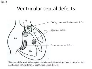
Embryology • Failure of the bulbar ridges to form the membranous part of the septum result in the most common forms of V.S.D. . • Defects of the muscular septum are usually ventral & high near the aortic orifice ; only rarely do they occur in the neighborhood of the apex
Pathophysiology • The physical size of the defect is a major, but not the only , determinant of the size of the left-to-right shunt . • The shunt magnetite is also determined by the level of pulmonary vascular resistance compared with systemic vascular resistance.
Pathophysiology • When a small communication is present (usually < 0.5 cm 2), the defect is called restrictive & right ventricular pressure is normal . • The higher pressure in the left ventricle drives the shunt left-to right. • In large non-restrictive defects (usually > 1.0 cm2), right & left ventricular pressures are equalized.
Pathophysiology • In these defects, the direction of shunting & the shunt magnitude are determined by the ratio of pulmonary to systemic vascular resistances. • After birth, in the presence of a large V.S.D.,the pulmonary vascular resistance may remain higher than normal & thus the size of the left-to-right shunt maybe limited . • As pulmonary vascular resistance falls in the 1st few weeks after birth because of the normal involution of the media of the small pulmonary Arteries & arterioles,the size of the left-to-right shunt increases & clinical symptoms become apparent.
Pathophysiology • When the ratio of pulmonary to systemic resistance approaches 1:1, the shunt becomes bi-directional, signs of heart failure abate (diminished), & the patient becomes cyanotic (Eisenmenger physiology). • Prolonged pulmonary hypertension is prevented by early surgical intervention in patients with large V.S.D.s
Pathophysiology • The magnitude of intracardiac shunts is usually described by the ratio of pulmonary to systemic blood flow. • If the left-to-right shunt is small (pulm. to syst. flow ratio <1.75:1), the cardiac chambers will not be appreciably enlarged & the pulm. Vascular bed will likely be normal. • If the shunt is large (flow ratio >2.5:1), the left atrial & ventricular volume overloaded occur, as well as right ventricular & pulm. Arterial hypertension. • The pulm. Arterial trunk, left atrium, & left ventricle are enlarged because of the large volume of pulmonary blood flow.
Asymptomatic. Cardiac lesion is usually found via a routine physical exam. (harsh ,loud ,blowing,left parasternal holosystolic murmer on left lower sternal border . The systolic murmer maybe not audible via the 1st few days(limited left-to-right shunt d.t.higher right side pr. CXR usually normal . ECG usually normal but may suggest LVH (RVH suggest pulmonary HTN , large VSD ,or associated symptom as pulmonary Stenosis) Clinical Manifestationssmall defects with trivial left-to-right shunts & NL. Pulm. Art. Pr.
Dyspnea , feeding difficulties , poor growth , recurrent pulmonary infections & cardiac failure in early infancy (cyanosis is usually absent ;but duskiness is noted during infections or crying. A palpable parasternal lift , a holosystolic murmer , less harsh & more blowing d.t. absence of a significant pr. Gradient across the defect. CXR showed increase broncho-vascular markings ,gross cardomegaly with prominence of both ventricles , left atrium & pulmonary artery, frank pulm. edema & pleural effusion. ECG shows bi-ventricular hypertrophy , P wave maybe notched or peaked. Clinical Manifestationslarge defects with excessive pulmonary blood flow & pulmonary HTN
Diagnosis • Two-dimensional echocardiogram: • position & size of VSD. • By Doppler examination used to diagnose very small muscular septum defect & the degree of volume overload of the left atrium & left ventricle. • Pulsed Doppler calculate the pr. Gradient across the defect.this will allow estimation of right ventricular pr. & help to determine whether the patient is at risk for the development of early pulmonary vascular disease.
Diagnosis • Cardiac catheterization • indicated whenclinical evaluation leaves uncertainly regarding the size of the shunt or when lab. data don’t fit well with the clinical findings. • It is useful for detecting the presence of associated cardiac defects.
Prognosis & Complications • (30-50%) of small VSDs will close spontaneously , most frequently during the 1st year of life • the vast majority of defects that close will do so before age 4 yr.. • One of the long term risks for these patients is that of infective endocarditis.
Prognosis & Complications • Endocarditis occurs in fewer than 2% of children with VSD , is more common in adolescents ,& is rare in children under 2 yr. of age • it is less common for moderate or large VSD to close spontaneously , even defects large enough to result in H.F.(manifested in infants as F.T.T.) may become smaller& rarely will close completely. • Large defects ass. with recurrent chest infections & C.H.F.
Treatment(small defects) • Reassure parents • allow the child to live a normal life • surgical repair is not recommended • protection against infective endocarditis • follow up screening for pulmonary HTN or pulmonic Stenosis indicated by RVH.
Treatment (large VSD) • Medical treatment has two aims : to control CHF & to prevent development of pulmonary vascular disease. • Patients show signs of recurrent chest infections & FTT. • Pulmonary vascular dse is prevented when surgery is performed within the 1st yr. of life. • Large defects ass. With pulmonary HTN should be closed between 6 & 12 mo. of age.
Treatment (large VSD) • Surgical risks are higher for defects in the muscular septum , particularly apical defects & multiple (Swiss-cheese type) defects,they may require pulmonary artery binding. • Catheter occlusion devices are currently being tested to close apical muscular VSDs. • After obliteration of the left-to-right shunt,catch-up growth occurs in the majority over the next 1-2 yr. • Systolic ejection murmurs of low intensity may persist for months.