Download
1 / 19
190 likes | 214 Views
Explore the complex structures of the facial skeleton, focusing on the orbit, its walls, connections, and variations. Learn about key components such as supraorbital margin, neurocranium, and fossae. Discover insights on the intricate facial bones and their contributions to the overall cranial framework.

E N D
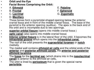
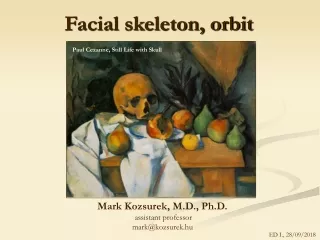
Facial skeleton, orbit Paul Cezanne, Still Life with Skull Mark Kozsurek, M.D., Ph.D. assistant professor mark@kozsurek.hu ED I., 28/09/2018
Supraorbital margin NEUROCRANIUM FACIAL SKELETON External acustic meatus
+ infratemporal fossa pterygopalatine fossa
Paired: • zygomatic • maxilla • nasal • palatine • lacrimal • inferior nasal concha Unpaired: • sphenoid • ethmoid • vomer also contribute to the neurocranium!
temporal fossa Zygomatic bone Surfaces: - orbital - lateral - temporal Processes: - frontal - maxillary - temporal Zygomatic canal: a Y-shaped channel with zygomaticoorbital, zygomaticfacial and zygomaticotemporal foramina
medial wall lateral wall inferior wall
Superior: • orbital part of the frontal • lesser wing of the sphenoid Medial: • frontal process of the maxilla • lacrimal • orbital lamina of the ethmoid • body of the sphenoid Lateral: • orbital surface of the zygomatic • orbital surface of the greater wing of the sphenoid Inferior: • orbital surface of the maxilla • orbital process of the palatine
1. aditus orbitae 2. frontal notch 3 supraorbital foramen 4. zygomaticoorbital, zygomaticofacial and zygomaticotemporal foramina (zygomatic canal) 5. infraorbital sulcus, canal and foramen 6. nasolacrimal canal 7. ant. ethmoidal foramen and canal 8. post. ethmoidal foramen and canal 9. optic canal 10. sup. orbital fissure 11. inf. orbital fissure 3 2 8 7 9 1 10 6 11 4 5
7. ant. ethmoidal foramen and canal 8. post. ethmoidal foramen and canal 9. optic canal 10. superior orbital fissure 7 8 9 10
11. inferior orbital fissure 11 Pterygopalatine fossa (deep inside) Infratemporal fossa
Structures within the orbit related to the lacrimal apparatus trochlear pit/spine fossa for lacrimal gland fossa for lacrimal sac
supraorbital notch and frontal foramen nose supraorbital notch and frontal notch supraorbital notch only nose nose supraorbital foramen and frontal foramen nose
"Blow-out" fracture of the orbit The eye and socket is hit by a fast moving object like a ball or fist. Usually the inferior and medial walls are involved. This can cause the eye to look sunken in or even cause double vision.