Download
1 / 36
360 likes | 374 Views
Immune System. Innate Defenses. External defenses. Skin Secretions. pH=3-5. Lysozyme Tears mucus, saliva. Mucus Membranes. Internal Innate Defense: Phagocytosis. Phagocytic cells.

E N D
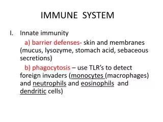
Innate Defenses External defenses • Skin • Secretions pH=3-5 • Lysozyme • Tears • mucus, • saliva
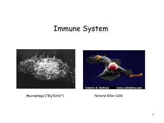
Internal Innate Defense: Phagocytosis Phagocytic cells Migrate OUT of the blood when the sense differences in concentration of certain chemicals engulf bacteria, dead cells, etc….
Natural Killer Cells Recognize surface molecules on abnormal cells (cancerous or virus infected)
Inflammatory response Swelling Pin Skin surface Bacteria Phagocytes and fluid move into area Phagocytes Chemical signals White blood cell Blood vessel Tissue injury; release of chemical signals such as histamine Phagocytes (macrophages and neutrophils) consume bacteria and cell debris; tissue heals Dilation and increased leakiness of local blood vessels; migration of phagocytes to the area 2 3 1 link
Pin Skin surface Fig. 24-2a Bacteria Chemical signals White blood cell Blood vessel Tissue injury; release of chemical signals such as histamine 1
Swelling Phagocytes and fluid move into area Fig. 24-2b Dilation and increased leakiness of local blood vessels; migration of phagocytes to the area 2
Phagocytes Fig. 24-2c Phagocytes (macrophages and neutrophils) consume bacteria and cell debris; tissue heals 3
Lymphatic system includes: -vessels (with valves) -fluid (lymph) -organs Important cells involved are T lymphocytes and B lymphocytes These cells are responsible for specific immune responses to specific pathogens
Acquired Immunity (the immune response) • Is highly specific • Produces antibodies in response to specific antigens • Antigens may be molecules on bacteria, viruses, protozoa, worms, transplanted organs • Both B and T lymphocytes have receptors on membrane that recognize different antigens
B cells -mature in bone - produce antibodies -antibodies float through the blood, recognizing and attaching to antigens T cells -mature in thymus -do not produce antibodies -cytotoxic T cells - require cell/cell contact to destroy pathogen Both B cells and T cells can produce memory cells
Cell-mediated immune response Humoral immune response Bone marrow Stem cell Thymus Via blood Fig. 24-5a Immature lymphocytes Antigen receptor Antigen receptor B cell T cell Via blood Lymph nodes, spleen, and other lymphatic organs Final maturation of B and T cells in lymphatic organ
Cell-mediated immune response Humoral immune response Bone marrow Stem cell Thymus Via blood Fig. 24-5a Immature lymphocytes Antigen receptor Antigen receptor B cell T cell Via blood Lymph nodes, spleen, and other lymphatic organs Final maturation of B and T cells in lymphatic organ
Primary Immune Response T cells are selected B cells are selected -antibody producing plasma cells are produced Person feels ill while these cells are produced Symptoms diminish as these cells do their job
Secondary Immune Response Response is much faster Memory cells are activated -tend to have a stronger response than the primary
Primary immune response Antigen receptor (antibody on cell surface) 1 B cells with different antigen receptors Fig. 24-7aa-1
Primary immune response 2 Antigen molecules Antigen receptor (antibody on cell surface) 1 B cells with different antigen receptors Fig. 24-7aa-2
Primary immune response 2 Antigen molecules Antigen receptor (antibody on cell surface) 1 B cells with different antigen receptors Fig. 24-7aa-3 First exposure to antigen 3 Cell activation: growth, division, and differentiation
Primary immune response 2 Antigen molecules Antigen receptor (antibody on cell surface) 1 B cells with different antigen receptors Fig. 24-7aa-4 First exposure to antigen 3 Cell activation: growth, division, and differentiation Antibody molecules 4 Endoplasmic reticulum First clone Plasma (effector) cells secreting antibodies
Primary immune response 2 Antigen molecules Antigen receptor (antibody on cell surface) 1 B cells with different antigen receptors Fig. 24-7aa-5 First exposure to antigen 3 Cell activation: growth, division, and differentiation Antibody molecules 4 5 Endoplasmic reticulum First clone Plasma (effector) cells secreting antibodies Memory cells
Antigen molecules Second exposure to same antigen 6 Fig. 24-7aa-6 Antibody molecules Secondary immune response (May occur long after primary immune response.) Endoplasmic reticulum Second clone Plasma (effector) cells secreting antibodies Memory cells
T cells work by directly binding to infected cells and then destroying them Cytotoxic T cell binds to infected cell 1 Self-nonself complex Fig. 24-12-1 Foreign antigen Infected cell Perforin molecule Cytotoxic T cell
Perforin makes holes in infected cell’s membrane and enzyme enters Cytotoxic T cell binds to infected cell 2 1 Self-nonself complex Fig. 24-12-2 Hole forming Foreign antigen Infected cell Perforin molecule Cytotoxic T cell
Infected cell is destroyed Perforin makes holes in infected cell’s membrane and enzyme enters Cytotoxic T cell binds to infected cell 2 3 1 Self-nonself complex Fig. 24-12-3 Hole forming Foreign antigen Infected cell Perforin molecule Cytotoxic T cell
Allergies Hypersensitivity to environmental antigen (allergen) Antibodies attach to mast cells - later, allergen attaches to these antibodies on mast cells Histamine & other inflammatory agents released
B cell (plasma cell) Fig. 24-17a Mast cell Antigenic determinant Histamine 2 Antibodies attach to mast cell Allergen (pollen grain) enters bloodstream B cells make antibodies 1 3 Sensitization: Initial exposure to allergen
Fig. 24-17b Histamine is released, causing allergy symptoms Allergen binds to antibodies on mast cell 4 5 Later exposure to same allergen
Anaphylactic shock Acute reaction to allergen Massive dilation of blood vessels -drop in blood pressure Counteracted by epinephrine
Active immunity results from natural recovery from infections vaccinations Passive immunity Receive antibodies from someone else -IgG anitibodies cross placenta -breast milk -shots (rabies treatment)
Transfusions/transplants ABO blood group -IgM doesn’t cross placenta Antibodies produced against bacterial antigens which are very similar rH factor -IgG crosses placenta
Tissue graphs/ organ transplants Give drugs that suppress cell mediated immunity Bone marrow transplants Risk of graft vs host reaction Donor lymphocytes attack host cells
Autoimmune diseases Immune system doesn’t recognize “self” and attacks MS Insulin dependent diabetes
AIDS HIV infection of cells require CD4 -found on T cells Is a retrovirus Antibodies are ineffective because -provirus gives it “invisibility” -rapid rate of mutation -Helper T cells decrease -secondary infections Drug treatments slow viral replication -AZT (reverse transcriptase inhibitors) -protease inhibitors