Fluid and Electrolyte Homeostasis
440 likes | 1.51k Views
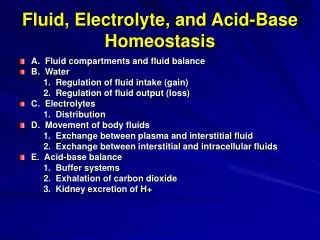
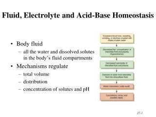
Fluid and Electrolyte Homeostasis. Na + and water: ECF volume and osmolarity K + : cardiac and muscle function Ca 2+ : exocytosis, muscle contractions, and other functions H + and HCO 3 – : pH balance Body must maintain mass balance Excretion routes: kidney and lungs.

Fluid and Electrolyte Homeostasis
E N D
Presentation Transcript
Fluid and Electrolyte Homeostasis • Na+ and water: ECF volume and osmolarity • K+: cardiac and muscle function • Ca2+: exocytosis, muscle contractions, and other functions • H+ and HCO3–: pH balance • Body must maintain mass balance • Excretion routes:kidney and lungs
Water Balance in the Body Figure 20-2
Water Balance A model of the role of the kidneys in water balance Figure 20-3
Fluid and Electrolyte Homeostasis The body’s integrated response to changes in blood volume and blood pressure Figure 20-1a
Fluid and Electrolyte Homeostasis Figure 20-1b
Urine Concentration Osmolarity changes as filtrate flows through the nephron Figure 20-4
Water Reabsorption Water movement in the collecting duct in the presence and absence of vasopressin Figure 20-5a
Water Reabsorption Figure 20-5b
Water Reabsorption Cross-section of kidney tubule Medullary interstitial fluid Collecting duct lumen Vasa recta Collecting duct cell 600 mOsM Filtrate 300 mOsm 600 mOsM 700 mOsM 1 Vasopressin Vasopressin receptor Vasopressin binds to mem- brane receptor. 1 The mechanism of vasopressin action Figure 20-6, step 1
Water Reabsorption Cross-section of kidney tubule Medullary interstitial fluid Collecting duct lumen Vasa recta Collecting duct cell 600 mOsM Filtrate 300 mOsm 600 mOsM 700 mOsM Second messenger signal 2 1 Vasopressin cAMP Vasopressin receptor Vasopressin binds to mem- brane receptor. Receptor activates cAMP second messenger system. 1 2 Figure 20-6, steps 1–2
Water Reabsorption Cross-section of kidney tubule Medullary interstitial fluid Collecting duct lumen Vasa recta Collecting duct cell 600 mOsM Filtrate 300 mOsm 600 mOsM 700 mOsM Storage vesicles Second messenger signal 2 Exocytosis of vesicles 1 Vasopressin Aquaporin-2 water pores cAMP 3 Vasopressin receptor Cell inserts AQP2 water pores into apical membrane. Vasopressin binds to mem- brane receptor. Receptor activates cAMP second messenger system. 1 2 3 Figure 20-6, steps 1–3
Water Reabsorption Cross-section of kidney tubule Medullary interstitial fluid Collecting duct lumen Vasa recta Collecting duct cell 600 mOsM Filtrate 300 mOsm H2O 600 mOsM H2O H2O H2O 700 mOsM 4 Storage vesicles Second messenger signal 2 Exocytosis of vesicles 1 Vasopressin Aquaporin-2 water pores cAMP 3 Vasopressin receptor Vasopressin binds to mem- brane receptor. Receptor activates cAMP second messenger system. Cell inserts AQP2 water pores into apical membrane. Water is absorbed by osmosis into the blood. 4 1 2 3 Figure 20-6, steps 1–4
Factors Affecting Vasopressin Release Main Trigger Figure 20-7
Water Balance The effect of plasma osmolarity on vasopressin secretion by the posterior pituitary Figure 20-8
Countercurrent Heat Exchanger Figure 20-9
Water Balance Countercurrent exchange in the medulla of the kidney Figure 20-10
Ion reabsorption “Loop Diuretics” furosemide (Lasix) x Active reabsorption of ions in the thick ascending limb creates a dilute filtrate in the lumen Figure 20-11
Fluid and Electrolyte Balance • Vasa recta removes water • Close anatomical association of the loop of Henle and the vasa recta--“countercurrent exchange” • Urea increases the osmolarity of the medullary interstitium--urea is 50% of solutes
Sodium Balance Homeostatic responses to salt ingestion Figure 20-12
Sodium Balance Aldosterone combines with a cytoplasmic receptor. 1 Interstitial fluid P cell of distal nephron Blood 2 Hormone-receptor complex initiates transcription in the nucleus. Lumen of distal tubule Transcription Aldosterone 2 1 mRNA Translation and protein synthesis 3 Aldosterone receptor 3 New protein channels and pumps are made. New channels New pumps ATP 4 Proteins modulate existing channels and pumps. Aldosterone- induced proteins modify existing proteins. 4 K+ secreted K+ K+ K+ 5 ATP Na+ reabsorbed Na+ Na+ Result is increased Na+ reabsorption and K+ secretion. 5 Na+ Aldosterone action in principle cells Figure 20-13
Sodium Balance The renin-angiotensin-aldosterone pathway Figure 20-14
Sodium Balance Decreased blood pressure stimulates renin secretion Figure 20-15
Sodium Balance Action of natriuretic peptides Figure 20-16
Potassium Balance • Regulatory mechanisms keep plasma potassium in narrow range • Aldosterone plays a critical role (mentioned earlier) • Hypokalemia • Muscle weakness and failure of respiratory muscles and the heart • Hyperkalemia • Can lead to cardiac arrhythmias • Causes include kidney disease, diarrhea, and diuretics
Disturbances in Volume and Osmolarity Figure 20-17
Volume and Osmolarity Blood volume/ Blood pressure DEHYDRATION CARDIOVASCULAR MECHANISMS Carotid and aortic baroreceptors CVCC Sympathetic output Parasympathetic output Heart Arterioles Vasoconstriction Rate Force Peripheral resistance Cardiac output Blood pressure Figure 20-18 (1 of 6)
Volume and Osmolarity Blood volume/ Blood pressure Osmolarity accompanied by DEHYDRATION HYPOTHALAMIC MECHANISMS Hypothalamic osmoreceptors Atrial volume receptors; carotid and aortic baroreceptors Hypothalamus + Vasopressin release from posterior pituitary + Thirst Distal nephron H2O intake H2O reabsorption Blood pressure Volume Osmolarity Figure 20-18 (2 of 6)
Volume and Osmolarity Blood volume/ Blood pressure DEHYDRATION RENIN-ANGIOTENSIN SYSTEM RENAL MECHANISMS + + Flow at macula densa Granular cells GFR Volume conserved Renin Angiotensinogen ANG I ACE ANG II Figure 20-18 (3 of 6)
Volume and Osmolarity Blood volume/ Blood pressure Osmolarity accompanied by DEHYDRATION RENIN-ANGIOTENSIN SYSTEM + Granular cells CVCC + Renin Angiotensinogen ANG I Vasopressin release from posterior pituitary ACE + + + Arterioles ANG II Thirst + osmolarity inhibits Adrenal cortex Aldosterone Distal nephron Na+ reabsorption Figure 20-18 (4 of 6)
Volume and Osmolarity Blood volume/ Blood pressure Osmolarity accompanied by DEHYDRATION CARDIOVASCULAR MECHANISMS RENIN-ANGIOTENSIN SYSTEM RENAL MECHANISMS HYPOTHALAMIC MECHANISMS Hypothalamic osmoreceptors + Atrial volume receptors; carotid and aortic baroreceptors Carotid and aortic baroreceptors + + Flow at macula densa Granular cells GFR + CVCC + Hypothalamus Volume conserved Renin + Angiotensinogen ANG I Vasopressin release from posterior pituitary Sympathetic output Parasympathetic output ACE + + + + Heart Arterioles ANG II Thirst + osmolarity inhibits Adrenal cortex Vasoconstriction Rate Force Aldosterone Peripheral resistance Distal nephron Distal nephron Na+ reabsorption H2O intake H2O reabsorption Cardiac output Blood pressure Volume Osmolarity Figure 20-18 (6 of 6)