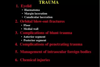
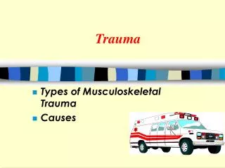
Trauma
Trauma. Types of Musculoskeletal Trauma Causes. Emergency Care. ABC’s Musculoskeletal Trauma Assess Circulation Immobilize. First Aid Keys!. ABCs First CMS Immobilize RICE. Pain!. Sprains, Strains. Strains ( tendons,muscle) Acute Chronic Classification 1st degree 2nd degree

Trauma
E N D
Presentation Transcript
Trauma Types of Musculoskeletal Trauma Causes
Emergency Care • ABC’s • Musculoskeletal Trauma • Assess • Circulation • Immobilize
First Aid Keys! ABCs First CMS Immobilize RICE
Pain! Sprains, Strains • Strains (tendons,muscle) • Acute • Chronic • Classification • 1st degree • 2nd degree • 3rd degree
What kind of injury? Sprain or strain?
Ligamentous Injury Sites Classification 1st degree 2nd degree 3rd degree Sprains
Nursing Diagnosis Immediate Care Rice Interventions Grade I=RICE Grade II Strain Sprain Grade III Management Sprains &Strains
Knee InjuriesTypesTerrible trial (medial collateral, anterior cruciate, medial meniscus) AssessmentManagement
Definitions Assessment Nursing diagnosis Examples Management Reduce Stabilize Slings Velpeau bandage Immobilizer Splints Dislocations/Subluxations
Hips also dislocate. In what positions can a hip dislocate? How do you detect hip dislocation in an infant?
Signs and symptoms of hip dislocation: anterior and posterior
Your patient presents in the ER with her hand wrapped in a cloth. What assessment would you make? What actions should you take?
Classifications Open Closed, complete Incomplete Impacted Comminuted Displaced Complicated By Direction Transverse Oblique Spiral Angulated Offset By Force By Physician Joint Injury Fractures
Significance of Fracture Description • Why? • Examples of Fracture • Angulated • Communiuted • Oblique • Stress Fx • Compression
Test Your Skill! Describe these fractures!
Fractures of the epiphyseal plate. What potential injury may result which has long term implications? Stress fracture Salter Fractures (epiphyseal plate fractures)
Steps in Bone Healing • Inflammation • Cellular Proliferation • Callus Formation • Ossification • Consolidation and Remodeling
Identify the steps in the bone healing process. What factors affect bone healing?
These slides reflect bone healing in a young person. Determine what stage of healing is reflected in each picture.
Influenced by Approximation of Bone Ends Circulation Nutrition Type of bone Nursing diagnosis Fracture Management Examples of Fractures Bone Healing Promoted by:
JR, 44 yr. old male, fell 12 feet; sustained open communiuted l. tibial fx. and closed spiral fx midshaft l. femur. Hx. 1 qt Wild Turkey daily. Wt. 240 H&H: 12; 36%; K:3.0; albumin: 2.5g/dl; amylase: 260 IU/L; T 102; FBS 150. Clinical Situation List at least 7 factors that will NEGATIVELY effect JR’s bone healing.
Circulation first Deformity Ecchymosis Muscle spasm Pain Abnormal movement Crepitus FractureAssessment/management
Colles: fx distal radius Open fx radius Supracondylar fx humerus: use Dunlop’s traction Shaft of humerus; use sling and swath Clavicle fx: use fig. 8 or clavicle strap Pelvic fx: injuries to bladder, iliac artery, sciatic nerve Open fx femoral shaft: blood loss Communiuted fx. distal 1/3 tibia Common Fractures and Management
Colles Fracture Produces a “silverfork” deformity
What observations? What assessment is most critical?
Describe this fracture. What is the anatomical location of the fracture? What type of traction might initially be used? A potential result of this fracture is Volksman’s ischemic contracture due to compartment syndrome.
Type of traction to initially treat a supracondylar fracture of the humerous
Fracture of the mid-shaft of the Humerus What potential complications might occur?
Fracture of the mid 1/3 of the clavicle Treatment with figure of 8 or clavicle strap. What teaching should be done?
Pelvic fractures may be treated surgically or with a pelvic fixator. Pelvis fractures vary in severity. What potential serious injuries may result form a pelvic fracture?
What method of treatment has been selected for this person with a pelvic fracture?
Describe this fracture. What are two major concerns? Treatment options: traction, ORIF
Describe the fracture including location. Why might this site require grafting?