TRAUMA
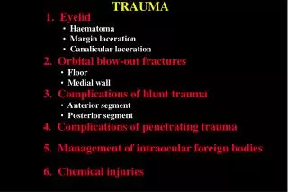
TRAUMA. 1. Eyelid. Haematoma Margin laceration Canalicular laceration. 2. Orbital blow-out fractures. Floor Medial wall. 3. Complications of blunt trauma. Anterior segment Posterior segment. 4. Complications of penetrating trauma.

TRAUMA
E N D
Presentation Transcript
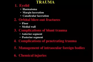
TRAUMA 1. Eyelid • Haematoma • Margin laceration • Canalicular laceration 2. Orbital blow-out fractures • Floor • Medial wall 3. Complications of blunt trauma • Anterior segment • Posterior segment 4. Complications of penetrating trauma 5. Management of intraocular foreign bodies 6. Chemical injuries
Eyelid haematoma Usually innocuous but exclude associated trauma to globe or orbit Orbital roof fracture if associated with subconjunctival haemorrhage without visible posterior limit Basal skull fracture - bilateral ring haematomas (‘panda eyes’)
Lid margin laceration Carefully align to prevent notching Align with 6-0 black silk suture Close tarsal plate with fine absorbable suture Close skin with multiple interrupted 6-0 black silk sutures Place additional marginal silk sutures
Canalicular laceration • Locate and approximate ends of laceration • Bridge defect with silicone tubing • Leave in situ for about 3 months • Repair within 24 hours
Signs of orbital floor blow-out fracture • Enophthalmos - if severe • Periocular ecchymosis • and oedema • Infraorbital nerve • anaesthesia • Ophthalmoplegia - • typically in up- and down- • gaze (double diplopia)
Investigations of orbital floor blow-out fracture Coronal CT scan Hess test • Restriction of right upgaze and downgaze • Secondary overaction of left eye • Right blow-out fracture with • ‘tear-drop’ sign
Surgical treatment of blow-out fracture a b c d (a) Subciliary incision • Coronal CT scan following repair of • right blow-out fracture with synthetic • material (b) Periosteum elevated and entrapped orbital contents freed (c) Defect repaired with synthetic material (d) Periosteum sutured
Medial wall blow-out fracture Signs Periorbital subcutaneous emphysema Ophthalmoplegia - adduction and abduction if medial rectus muscle is entrapped Treatment • Release of entrapped tissue • Repair of bony defect
Anterior segment complications of blunt trauma Vossius ring Hyphaema Iridodialysis Sphincter tear Cataract Lens subluxation Angle recession Rupture of globe
Posterior segment complications of blunt trauma Choroidal rupture and haemorrhage Avulsion of vitreous base and retinal dialysis Commotio retinae Macular hole Optic neuropathy Equatorial tears
Complications of penetrating trauma Uveal prolapse Damage to lens and iris Flat anterior chamber Vitreous haemorrhage Tractional retinal detachment Endophthalmitis
Management of intraocular foreign bodies Removal with magnet or by pars plana vitrectomy Localization with reference to radio- opaque marker
Grading of severity of chemical injuries Grade I (excellent prognosis) • Clear cornea • Limbal ischaemia - nil Grade III (guarded prognosis) Grade IV (very poor prognosis) Grade II (good prognosis) • No iris details • Opaque cornea • Cornea hazy but visible • iris details • Limbal ischaemia > 1/2 • Limbal ischaemia - 1/3 to 1/2 • Limbal ischaemia < 1/3
Medical Treatment of Severe Injuries 1. Copious irrigation ( 15-30 min ) - to restore normal pH 2. Topical steroids ( first 7-10 days ) - to reduce inflammation 3. Topical and systemic ascorbic acid - to enhance collagen production 4. Topical citric acid - to inhibit neutrophil activity 5. Topical and systemic tetracycline - to inhibit collagenase and neutrophil activity
Surgical treatment of severe chemical injuries Division of conjunctival bands Treatment of corneal opacity by keratoplasty or keratoprosthesis Correction of eyelid deformities