Download

1 / 39
390 likes | 565 Views
Osteomyelitis in Children. Dr. Robert Deane Janeway. Age Incidence Etiology Pathophysiology Presentation Laboratory investigations Imaging. Treatment Surgery Complications Summary Special Groups. Outline. Age / Incidence / Etiology. 1/1000 – 1/ 20 000 Male > Female

E N D
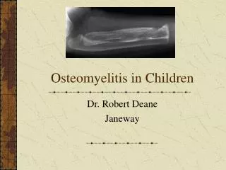
Osteomyelitis in Children Dr. Robert Deane Janeway
Age Incidence Etiology Pathophysiology Presentation Laboratory investigations Imaging Treatment Surgery Complications Summary Special Groups Outline
Age / Incidence / Etiology • 1/1000 – 1/ 20 000 • Male > Female • Pre antibiotic era ……20-50% mortality
Age / Incidence / Etiology • Advances in treatment • Earlier dx • Antibiotic tx • Surgery less delay • Children better nourished
Age / Incidence / Etiology • Glasgow incidence decreased • New Zealand……. Madri > Whites • South Africa…….. Black > Whites • Changing disease / Changing organism • Seasonal Variation • Nutritional status, climate, lifestyle
Age / Incidence / Etiology • H Flu • Big cause 1970’s • 1-4 yrs • Now decreased due to vaccinations • Kingella Kingae • OM in older kids • Septic Arthritis 1-3 yrs • Neonates separate group
Pathophysiology • Poorly defined • Direct inoculation • Hematogenous spread • Local invasion
Pathophysiology • Infection • Starts in Metaphysis • Arteriole Loop / Venous Lakes • Spread via Volkman’s canal / Haversian system • Endothelium Leaks
Pathophysiology • Few phagocytes in Zone of Hypertrophy • Highest incidence in fastest growing bone • Tubular > Flat bones
Pathophysiology • Gaps in endothelium metaphyseal vessel • Bacteria pass • Adhere to Type 1 collagen • Increase pressure in bone/ decrease blood flow • Bone infarction / Dead Bone (sequestrum)
Pathophysiology • Spread via Volkman Canal • Subperiosteal Pus • Cortex breaks down • May spread to joint • Hip / Shoulder / Fibula / Proximal Humerus
Pathophysiology • Role of Trauma • Rabbit experiment • IV injection of bacteria • With # start in hematoma
Pathophysiology • Role of growth plate • Over 18/12 • Impermeable to spread • Under 18/12 infection crosses growth plate
Pathophysiology • 1st osteoblasts die • Lymphocytes release osteoclast activating factor • Hole in bone
Diagnosis • Pain • Neonate peudoparalysis • NWB • Failure to use limb • Fever • Lethargy • Anorexia • Swelling (neonates / older kids)
Pathophysiology • Bloodwork • CBC Diff • ESR • CRP • Blood Culture
Pathophysiology • WBC increased 30-40% • Left Shift 65% • ESR increased 91%……….24-36hrs • CRP increased 97%…………4-6hrs
Pathophysiology • CRP • More rapid than ESR • 2-4 hrs …..peak 72hrs • 10-30x normal • Systemic ds (trauma, tumor)
Pathophysiology • Blood Culture • + 30-60% • Decreased with antibiotic • Multiple cultures no significant increase in yield • 48 hours to get most organisms
Diagnosis • Pus aspiration • 70% bone + cultures • Septic arthritis • Gram stain • Lymphocyte count • % polymorphs • > 80 000 = Septic arthritis • > 50 000 in some series • 80 000 also in JRA
Diagnosis • Do blood and joint cultures • One or other not always +ve in same pt • Gram stain +ve 1/3 bone and joint aspirations • Future looking for bacteria DNA / RNA
Lab Diagnosis • WBC not reliable • False sense of security • 25% increased Mayo clinic • 65% diff abnormal • Acute phase reactants • Change in plasma proteins d/t cytokines
Diagnosis • ESR • Nonspecific acute phase reactant • Depends on fibrinogen concentration • Increased 48-72 hrs • Increased in 90% of cases • Not affected by antibiotic tx • CRP • Increased in 98% of cases
Radiology • Plain xray • Sensitivity 43-75% • Specificity 75-83% • Soft tissue swelling 48hrs • Periosteal reaction 5-7d • Osteolysis 10d to 2 wks • (need 50% bone loss)
Radiology • Tc99 • 24-48hrs +ve • Bone aspiration DOES NOT give false +ve • Decreased uptake in early phase d/t increased pressure • “cold” scan up to 100% PPV
Radiology • Gallium • 48 hrs to do • Non specific • Indium • I131 leucocytes • 24hrs to prepare • Monoclonal antibodies • Not proven to be better
Radiology • MRI • Sensitivity 83-100% • Specificity 75-100% • PPV = Tc99 • Marrow and soft tissue swelling • Good in spine and pelvis
Radiology • T1 • Best for acute infection • Gadolinium helps • Changes similar to • # • Infarct • Bruise • Tumor • Post surgical • Sympathetic edema
Radiology • CT • Gas • sequestrum
Treatment • Mostly medical • Sx to improve local environment • Remove infected devitalized bone • Decompress abscess cavity • Timing !! • Early antibiotic before necrosis / pus then sx less likely to be needed
Treatment • Antibiotic treatment • Parenteral / oral combinations • Often empirical • Serum level more important than route • Follow WBC / ESR/ CRP • Organism / sensitivity
Treatment • Treatment Failure • High doses • Poor oral absorption / compliance • Inadequate monitoring of serum levels • Delay in Sx
Treatment • Previously start IV • Follow ESR to guide switch to oral • Newer studies • Follow CRP • Shorter period of tx needed • IV 5d / total 23 d tx • Cephalosporin 150mg/kd/day
Treatment • Neonates • No studies, little evidence • CRP / ESR not reliable • Oral absorption not reliable • Therefore IV neonates • Cloxacillin
Treatment • Longer treatment required • Pelvis • Vertebrae • Diskitis • Calcaneus
Treatment • Surgical intervention • Controversial indications • Hole in bone not always Sx • If purulent aspirate Sx necessary • Sx less frequent with newer antibiotic • 22-83% earlier studies • 8-43% recent studies
Treatment • Surgery Indicated • Subperiosteal Abscess • Soft Tissue abscess • Bone Abscess • Failure of clinical response to antibiotic • Associated septic arthritis
Complications • Infection Complications • Recurrence • Chronic osteo • Pathologic fracture • Growth plate injury • Antibiotic Complications • Diarrhea • N+V • Rash • Thrombocytopenia • Neutropenia