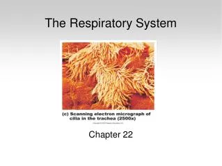
Chapter 22 Respiratory System
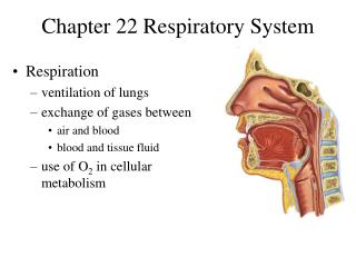
Chapter 22 Respiratory System. Respiratory System. Anatomy of the Respiratory System Pulmonary Ventilation Gas Exchange and Transport Respiratory Disorders. Organs of Respiratory System. Nose, pharynx, larynx, trachea, bronchi, lungs. General Aspects. Airflow in lungs

Chapter 22 Respiratory System
E N D
Presentation Transcript
Chapter 22 Respiratory System
Respiratory System • Anatomy of the Respiratory System • Pulmonary Ventilation • Gas Exchange and Transport • Respiratory Disorders
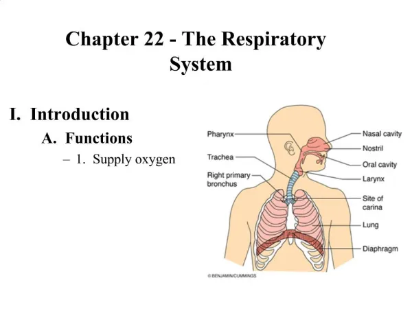
Organs of Respiratory System • Nose, pharynx, larynx, trachea, bronchi, lungs
General Aspects • Airflow in lungs • bronchi bronchioles alveoli • Conducting division • passages for airflow, nostrils to bronchioles • Respiratory division • distal gas-exchange regions, alveoli • Upper respiratory tract • organs in head and neck, nose through larynx • Lower respiratory tract • organs of thorax, trachea through lungs
Nose • Functions • warms, cleanses, humidifies inhaled air • detects odors • resonating chamber that amplifies the voice • Bony and cartilaginous supports • superior half: nasal bones medially and maxillae laterally • inferior half: lateral and alar cartilages • ala nasi: flared portion shaped by dense CT, forms lateral wall of each nostril
Nasal Cavity • Extends from nostrils to posterior nares • Vestibule: dilated chamber inside ala nasi • stratified squamous epithelium, vibrissae (guard hairs) • Nasal septum divides cavity into right and left chambers called nasal fossae
Nasal Cavity- Conchae and Meatuses • Superior, middle and inferior nasal conchae • 3 folds of tissue on lateral wall of nasal fossa • mucous membranes supported by thin scroll-like turbinate bones • Meatuses • narrow air passage beneath each conchae • narrowness and turbulence ensures air contacts mucous membranes
Nasal Cavity - Mucosa • Olfactory mucosa • lines roof of nasal fossa • Respiratory mucosa • lines rest of nasal cavity with ciliated pseudostratified epithelium • Defensive role of mucosa • mucus (from goblet cells) traps inhaled particles • bacteria destroyed by lysozyme
Nasal Cavity- Cilia and Erectile Tissue • Function of cilia of respiratory epithelium • sweep debris-laden mucus into pharynx to be swallowed • Erectile tissue of inferior concha • venous plexus that rhythmically engorges with blood and shifts flow of air from one side of fossa to the other once or twice an hour to prevent drying • Spontaneous epistaxis (nosebleed) • most common site is inferior concha
Pharynx • Nasopharynx (pseudostratified epithelium) • posterior to choanae, dorsal to soft palate • receives auditory tubes and contains pharyngeal tonsil • 90 downward turn traps large particles (>10m) • Oropharynx (stratified squamous epithelium) • space between soft palate and root of tongue, inferiorly as far as hyoid bone, contains palatine and lingual tonsils • Laryngopharynx (stratified squamous) • hyoid bone to level of cricoid cartilage
Larynx • Glottis – vocal cords and opening between • Epiglottis • flap of tissue that guards glottis, directs food and drink to esophagus • Infant larynx • higher in throat, forms a continuous airway from nasal cavity that allows breathing while swallowing • by age 2, more muscular tongue, forces larynx down
Nine Cartilages of Larynx • Epiglottic cartilage - most superior • Thyroid cartilage – largest; laryngeal prominence • Cricoid cartilage - connects larynx to trachea • Arytenoid cartilages (2) - posterior to thyroid cartilage • Corniculatecartilages (2) - attached to arytenoid cartilages like a pair of little horns • Cuneiformcartilages (2) - support soft tissue between arytenoids and epiglottis
Walls of Larynx • Interior wall has 2 folds on each side, from thyroid to arytenoid cartilages • vestibular folds: superior pair, close glottis during swallowing • vocal cords: produce sound • Intrinsic muscles - rotate corniculate and arytenoid cartilages • adducts (tightens: high pitch sound) or abducts (loosens: low pitch sound) vocal cords • Extrinsic muscles - connect larynx to hyoid bone, elevate larynx during swallowing
Trachea • Rigid tube 4.5 in. long and 2.5 in. diameter, anterior to esophagus • Supported by 16 to 20 C-shaped cartilaginous rings • opening in rings faces posteriorly towards esophagus • trachealis spans opening in rings, adjusts airflow by expanding or contracting • Larynx and trachea lined with ciliated pseudostratified epithelium which functions as mucociliary escalator
Bronchial Tree • Primary bronchi (C-shaped rings) • from trachea; after 2-3 cm enter hilum of lungs • right bronchus slightly wider and more vertical (aspiration) • Secondary (lobar) bronchi (overlapping plates) • one secondary bronchus for each lobe of lung • Tertiary (segmental) bronchi (overlapping plates) • 10 right, 8 left • bronchopulmonary segment: portion of lung supplied by each
Bronchial Tree • Bronchioles (lack cartilage) • layer of smooth muscle • pulmonary lobule • portion ventilated by one bronchiole • divides into 50 - 80 terminal bronchioles • ciliated; end of conducting division • respiratory bronchioles • divide into 2-10 alveolar ducts; end in alveolar sacs • Alveoli - bud from respiratory bronchioles, alveolar ducts and alveolar sacs • main site for gas exchange
Alveolus Fig. 22.11 b and c
Pleurae and Pleural Fluid • Visceral (on lungs) and parietal (lines rib cage) pleurae • Pleural cavity - space between pleurae, lubricated with fluid • Functions • reduce friction • create pressure gradient • lower pressure assists lung inflation • compartmentalization • prevents spread of infection
Pulmonary Ventilation • Breathing (pulmonary ventilation) – one cycle of inspiration and expiration • quiet respiration – at rest • forced respiration – during exercise • Flow of air in and out of lung requires a pressure difference between air pressure within lungs and outside body
Respiratory Muscles • Diaphragm (dome shaped) • contraction flattens diaphragm • Scalenes - hold first pair of ribs stationary • External and internal intercostals • stiffen thoracic cage; increases diameter • Pectoralis minor, sternocleidomastoid and erector spinae muscles • used in forced inspiration • Abdominals and latissimus dorsi • forced expiration (to sing, cough, sneeze)
Neural Control of Breathing • Breathing depends on repetitive stimuli from brain • Neurons in medulla oblongata and pons control unconscious breathing • Voluntary control provided by motor cortex • Inspiratory neurons: fire during inspiration • Expiratory neurons: fire during forced expiration • Fibers of phrenic nerve go to diaphragm; intercostal nerves to intercostal muscles
Respiratory Control Centers • Respiratory nuclei in medulla • inspiratory center (dorsal respiratory group) • frequent signals, you inhale deeply • signals of longer duration, breath is prolonged • expiratory center (ventral respiratory group) • involved in forced expiration • Pons • pneumotaxic center • sends continual inhibitory impulses to inspiratory center, as impulse frequency rises, breathe faster and shallower • apneustic center • prolongs inspiration, breathe slower and deeper
Input to Respiratory Centers • From limbic system and hypothalamus • respiratory effects of pain and emotion • From airways and lungs • irritant receptors in respiratory mucosa • stimulate vagal afferents to medulla, results in bronchoconstriction or coughing • stretch receptors in airways - inflation reflex • excessive inflation triggers reflex • stops inspiration • From chemoreceptors • monitor blood pH, CO2 and O2 levels
Chemoreceptors • Peripheral chemoreceptors • found in major blood vessels • aortic bodies • signals medulla by vagus nerves • carotid bodies • signals medulla by glossopharyngeal nerves • Central chemoreceptors • in medulla • primarily monitor pH of CSF
Voluntary Control • Neural pathways • motor cortex of frontal lobe of cerebrum sends impulses down corticospinal tracts to respiratory neurons in spinal cord, bypassing brainstem • Limitations on voluntary control • blood CO2 and O2 limits cause automatic respiration
Pressure and Flow • Atmospheric pressure drives respiration • 1 atmosphere (atm) = 760 mmHg • Intrapulmonary pressure and lung volume • pressure is inversely proportional to volume • for a given amount of gas, as volume , pressure and as volume , pressure • Pressure gradients • difference between atmospheric and intrapulmonary pressure • created by changes in volume thoracic cavity
Inspiration - Pressure Changes • intrapleural pressure • as volume of thoracic cavity ,visceral pleura clings to parietal pleura • intrapulmonary pressure • lungs expand with visceral pleura • Transpulmonary pressure • intrapleural minus intrapulmonary pressure (not all pressure change in the pleural cavity is transferred to the lungs) • Inflation aided by warming of inhaled air • 500 ml of air flows with a quiet breath
Passive Expiration • During quiet breathing, expiration achieved by elasticity of lungs and thoracic cage • As volume of thoracic cavity , intrapulmonary pressure and air is expelled • After inspiration, phrenic nerves continue to stimulate diaphragm to produce a braking action to elastic recoil
Forced Expiration • Internal intercostal muscles • depress the ribs • Contract abdominal muscles • intra-abdominal pressure forces diaphragm upward • pressure on thoracic cavity
Pneumothorax • Presence of air in pleural cavity • loss of negative intrapleural pressure allows lungs to recoil and collapse • Collapse of lung (or part of lung) is called atelectasis
Resistance to Airflow • Pulmonary compliance • distensibility of lungs; change in lung volume relative to a change in transpulmonary pressure • Bronchiolar diameter • primary control over resistance to airflow • bronchoconstriction • triggered by airborne irritants, cold air, parasympathetic stimulation, histamine • bronchodilation • sympathetic nerves, epinephrine
Alveolar Surface Tension • Thin film of water needed for gas exchange • creates surface tension that acts to collapse alveoli and distal bronchioles • Pulmonary surfactant (great alveolar cells) • decreasessurface tension • Premature infants that lack surfactant suffer from respiratory distress syndrome
Alveolar Ventilation • Dead air • fills conducting division of airway, cannot exchange gases • Anatomic dead space • conducting division of airway • Physiologic dead space • sum of anatomic dead space and any pathological alveolar dead space • Alveolar ventilation rate • air that ventilates alveoli X respiratory rate • directly relevant to ability to exchange gases
Measurements of Ventilation • Spirometer - measures ventilation • Respiratory volumes • tidal volume: volume of air in one quiet breath • inspiratory reserve volume • air in excess of tidal inspiration that can be inhaled with maximum effort • expiratory reserve volume • air in excess of tidal expiration that can be exhaled with maximum effort • residual volume (keeps alveoli inflated) • air remaining in lungs after maximum expiration