Download
1 / 20
200 likes | 556 Views
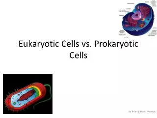
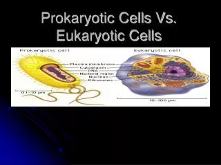
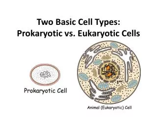
Prokayotic vs Eukaryotic Cells. Functional Anatomy. Typical Bacterial Cell. Typical Eukaryotic Cells. Prokaryote or “before nucleus” no membrane-bound nucleus no other membrane-bound organelles DNA not associated with histones cell walls almost always contain peptidoglycan 70s ribosomes

E N D
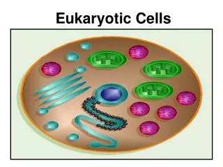
Prokayotic vs Eukaryotic Cells Functional Anatomy
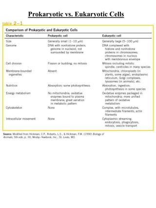
Prokaryote or “before nucleus” no membrane-bound nucleus no other membrane-bound organelles DNA not associated with histones cell walls almost always contain peptidoglycan 70s ribosomes Largest about size of smallest eukaryote Eukaryote or “true nucleus” membrane bound nucleus many other membrane-bound organelles DNA associated with histones cell walls never contain peptidoglycan 80s ribosomes Smallest about size of largest prokaryote Prokaryote vs Eukaryote Overview
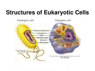
Prokaryotic Cells • Size • Smallest of living cells • 0.2 to 2.0 μm in diameter • 2 to 8 μm in length • Most eukaryotes bigger • Viruses much smaller
Cocci - spherical Bacilli – rods Spirillum - spiral Common Bacterial Shapes
Other, Less Common Shapes • Vibrio – comma • Coccobacillus - • Square • Star
Cocci Bacilli Common Cell arrangements
Prokaryotic Anatomy from the Outside In • Glycocalyx • Appendages • Cell Wall • Bacterial Cell Membranes • Inside the Cell
Glycocalyx • Sticky substances that surround cells • Firmly attached = capsule • Loosely attached = slime layer • Composition varies with species • Polysaccharides • Polypeptides • Both • Function • Protect cell from phagocytosis and dehydration • Aid in attachment to various surfaces • May inhibit movement of nutrients from cell
Appendages • Flagella • Tail-like structures extending out from glycocalyx • Functions in movement of the bacterial cell • Complex structure
Structure of Flagella • Filament • Long tail-like region • Constant diameter • Made of protein • Hook • Filament attachment • Basal body • Small central rod inserted into a series of rings
Cell Wall • Rigid • Composed mostly of peptidoglycan • Found only in bacterial cell walls • Amount differs in gram+ and gram- cells • Protects cell in environments with osmotic pressures
Glycan portion NAG N-acetylglucosamine NAM N-acetylmuramic acid Linked in rows of 10-65 sugars Peptide portion Adjacent rows are linked by polypeptides Peptidoglycan
Atypical Cell Walls • Mycoplasmas • Lack cell wall • Smallest known bacteria • Archeobacteria • Cell walls contain pseudomurein rather than peptidoglycan • Lacks D-amino acids found in bacteria • L-forms • Tiny mutant bacteria with defective cell walls • Just enough material to prevent lysis in dilute environments
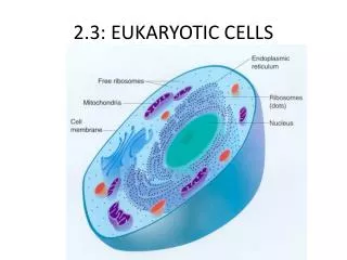
Inside the Cell Wall • Cell Membrane • Cytoplasm • 4/5 water and 1/5 dissolved substances • Most chemical reactions occur here • Ribosomes • Abundant in cytoplasm • 70s • Nuclear region • Central 10% of cell volume • DNA in single circular chromosome • Inclusions • small bodies within cytoplasm • Many different types