Understanding the Urinary System: Kidney to Bladder Connection
Explore the intricate details of the urinary system, from the bean-shaped kidneys to the bladder, with a focus on structures like the renal pelvis, nephrons, ureters, and urethra. Figures and descriptions help demystify the pathway of urine formation.


Understanding the Urinary System: Kidney to Bladder Connection
E N D
Presentation Transcript
URINARY SYSTEM STRUCTURE
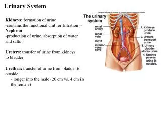
KIDNEY (FIGURE 20-2) • Bean-shaped • Located between peritoneum and the back muscles (retroperitoneal) • Renal pelvis – funnel-shaped structure at the beginning of the ureter • Medulla • Inner, striated layer • Striated cones are renal pyramids • Base of pyramids empty into cuplike cavities called calyces • Cortex – composed of millions of microscopic nephrons
NEPHRON (FIGURE 20-3) • Functional unit of kidney • Bowman’s capsule • Glomerulus • Proximal convoluted tubule • Loop of Henle • Distal convoluted tubule • Collecting tubule • See Figure 20-4 pg. 431 for “Pathway of urine formation” • Figure 20-5 pg. 432
URETERS • One from each kidney • Smooth muscle tube with mucous membrane lining
URINARY BLADDER • Hollow, muscular organ • Made of elastic fibers and involuntary muscle • Stores urine – about 500 cc
URETHRA • Connects bladder with urinary meatus • Urinary meatus is opening to body