Download
1 / 40
400 likes | 410 Views
Our aim is to alleviate human suffering related to diabetes and its complications among those least able to withstand the burden of the disease. From 2002 to March 2017, the World Diabetes Foundation provided USD 130 million in funding to 511 projects in 115 countries. For every dollar spent, the Foundation raises approximately 2 dollars in cash or as in-kind donations from other sources. The total value of the WDF project portfolio reached USD 377 million, excluding WDF’s own advocacy and strategic platforms.

E N D
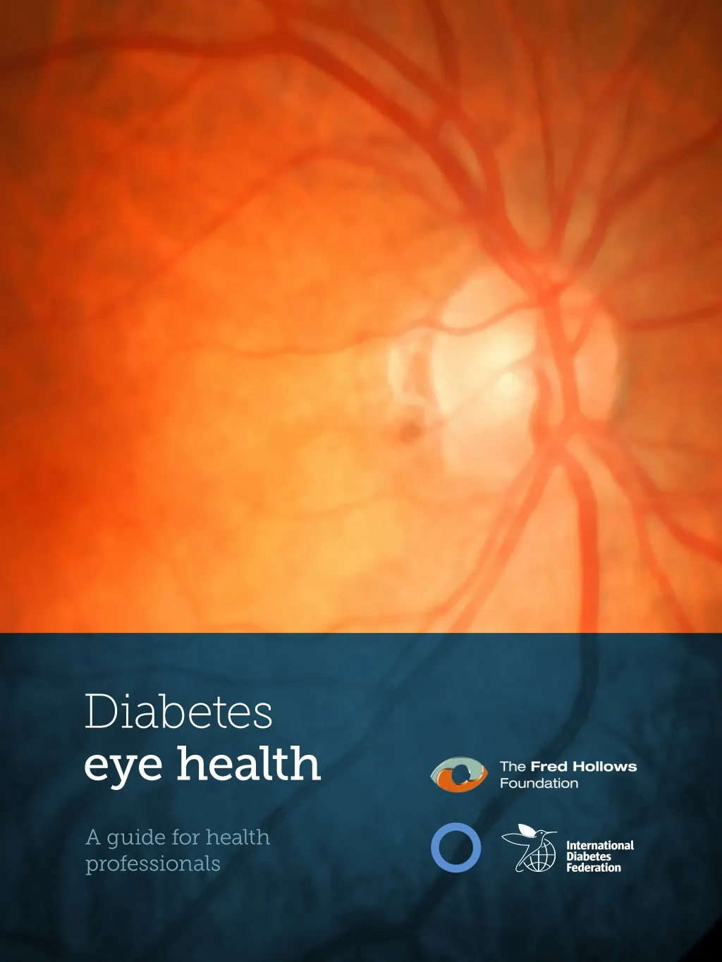
Diabetes eye health A guide for health professionals
Contributors A working group was convened to develop this Guide which included the following members: Co-Chairs: Sehnaz Karadeniz and Paul Zimmet Core Contributors: Pablo Aschner, Anne Belton, David Cavan, Atieno Jalang’o, Navleen Gandhi, Linda Hill, Lydia Makaroff, Richard Le Mesurier, Bina Patel, Massimo Porta, Hugh Taylor. The International Diabetes Federation and The Fred Hollows Foundation would also like to thank the following contributors: Haslina Binte Hamzah, Muhammad Daud Khan, Ute Linnenkamp, Vanessa Luttermann, Tim Nolan, Geneva Pritchard, Anna Saxby, Madeleine Smythe, Sara Webber, Wong Tien Yin. Support This publication was made possible with support from Bayer Pharma AG and Novartis Pharma AG. Published by the International Diabetes Federation © International Diabetes Federation and The Fred Hollows Foundation Retinal photographs are copyrighted by the Singapore Eye Research Institute. All Rights Reserved. The following photographs from the Community Eye Health Journal www.cehjournal.org are used under Creative Commons 2.0: “Screening and photo grading services” Indonesia. Photo: Dwi Ananta, HKI. “Participants in Trinidad, Tobago ‘walk for sight’ on World Sight Day 2013” Photo: IAPB/VISION 2020. “It is important to listen to the patient’s point of view” Bangladesh. Photo: Lutful Husain. “Patients wait for eye examination during community outreach” Democratic Republic of Congo. Photo: Daniel Etya’ale. “Examination of the eye” Mozambique. Photo: Riccardo Gangale/Sightsavers. “Ophthalmic staff preparing to see patients” Ethiopia. Photo Lance Bellers/Sight Savers. “Mobile unit” India. Photo: Project Nayantara. “A photographer working with a mobile clinic team takes fundus images in a rural hospital” Photo: Cristóvão Matsinhe. ISBN: 978-2-930229-82-9 Please cite this report as: International Diabetes Federation and The Fred Hollows Foundation. Diabetes eye health: A guide for health care professionals. Brussels, Belgium: International Diabetes Federation, 2015. www.idf.org/eyecare Cover image: Shutterstock © memorisz
Diabetes eye health Diabetic retinopathy affects over one third of all people with diabetes and is the leading cause of vision loss in working-age adults. Globally the prevalence of diabetes is increasing rapidly and without effective action, so too will the number of people with diabetic retinopathy. The International Diabetes Federation and The Fred Hollows Foundation have entered into a partnership to raise awareness of diabetic retinopathy. An outcome of the partnership has been to support a group of international experts to develop this Guide. We look forward to working with partners to promote the use of the Guide and ultimately to ensure people with diabetes have access to eye health services. The management of diabetes and its complications begins in primary health care and this should include screening for diabetic retinopathy. Those working in primary health care are at the frontline of supporting people with diabetes to understand how to look after their eyes, access eye health exams and to refer those requiring treatment. On behalf of our two organisations, we commend Diabetes eye health: A guide for health professionals. Sir Michael Hirst President International Diabetes Federation www.idf.org Mr Les Fallick Chair and The Fred Hollows Foundation www.hollows.org 3
Screening and photo grading services, Indonesia. Photo: Dwi Ananta, HKI. CC BY-NC 2.0 CEHJ 4
Table of contents Foreword 7 Executive summary 8 The purpose and scope of this document 9 What is diabetic eye disease? 10 Diabetes is increasing and so is diabetic eye disease 10 Managing diabetes to manage eye health 11 Keeping good eyes – the key players 12 The financial and social burden of eye disease 13 Identifying diabetic eye disease 14 Managing diabetes for good eye health 16 Different types of diabetes and implications for eye health 18 Strategies to managing eye health 19 Detection of diabetic retinopathy 20 Grading of diabetic retinopathy and macular edema 22 Ophthalmic assessment of diabetic eye disease 25 Treatment of diabetic retinopathy by ophthalmologists 26 Post-treatment support 29 Everyone with diabetes is at risk of diabetic retinopathy 29 Appendix 1: Managing Eye Health in People with Diabetes 30 Appendix 2: Managing Diabetes for good eye health 32 Glossary 34 References 36 Additional sources of information 38 5
Foreword for Diabetic Eye Care 2014 that set out the need for regular eye care from an ophthalmic perspective. The ICO guidelines stress the need for a team approach to the provision of care. This new Guide extends this approach to highlight what is needed from diabetologists, primary care practitioners and others involved in the care of people with diabetes. Everyone with diabetes is at risk of losing vision. Good control of blood glucose, blood pressure and blood lipids will reduce the annual incidence of eye disease and vision loss and will also prolong life. Timely treatment can prevent almost all vision loss associated with diabetes and so regular eye exams become essential for all those living with diabetes. Regular eye screening begins with primary health carers. There are only about 200,000 ophthalmologists worldwide and it would be impossible for them to undertake all the screening eye examinations required to detect those at risk of vision loss and in need of treatment. Screening for diabetic eye disease needs to become an integral part of the ongoing primary care of those with diabetes with the establishment of well-defined referral pathways for those needing further care. The ICO is delighted to see this collaborative approach to the provision of eye care for those people living with diabetes. We look forward to working with the International Diabetes Federation, The Fred Hollows Foundation, and others in the eye health and diabetes sectors, to promote the use of this Guide and to reduce the amount of blindness and vision loss from diabetes. Hugh R. Taylor AC MD President International Council of Ophthalmology This Guide builds on the guidelines developed by the International Council of Ophthalmology (ICO) Guidelines 7 Diabetes eye health – A guide for health professionals
Executive summary of people with diabetes, are most likely to have the opportunity to screen, educate and support management of diabetic eye disease. They can also facilitate timely referral to eye specialist services for treatment to reduce sight loss. More specialised eye health practitioners also have an important role however, as a relatively limited resource, they should focus on treatment rather than examination. The purpose of this document is to highlight for health professionals the rising prevalence of diabetic-related eye disease, particularly diabetic retinopathy, and outline the important role and actions they can take to address it. As the incidence of diabetes increases worldwide, so does the incidence of its complications including diabetic eye disease. All patients with diabetes are at risk of developing diabetic retinopathy. Diabetic retinopathy is the only eye condition caused by diabetes; however diabetes may exacerbate other eye conditions such as cataract, glaucoma, loss of focussing ability, and double vision. Key actions by health professionals in managing eye health in people with diabetes include: • Optimising control of blood glucose, blood pressure and blood lipids in order to slow down the progression of diabetic retinopathy • Ensuring that the person with diabetes has regular eye exams and timely treatment when required • Educating and supporting the person with diabetes in managing their eye health and their diabetes Diabetic retinopathy can cause blindness, yet in most cases blindness is largely avoidable. The condition is often asymptomatic in its early stages and regular eye examinations are the only way to determine the condition of the retina and take the appropriate action. Careful management of diabetes and early eye disease detection can help slow costly and debilitating visual impairment and blindness. Maintaining good vision requires optimising systemic factors (like blood glucose, blood pressure and blood lipid control), regular eye checks and timely referral for treatment. Effective strategies in managing diabetes to reduce or stabilise vision loss are through a combination of four key strategies: social support, nutritional support, medication, and medical examinations and treatment. The decision to undergo treatment should be made in cooperation between the person with diabetes and the health professional1. Primary health practitioners play a crucial role in all stages of managing good eye health by facilitating early diagnosis and timely management of diabetic eye disease. Many people with diabetes—as well as many health professionals—are unaware of the critical need to undergo regular eye examinations. Primary health professionals, through their routine care If diabetic retinopathy has been detected, referral to an ophthalmologist for timely treatment with laser photocoagulation and/ or intravitreal injections can prevent vision loss, stabilise vision, and in some cases even improve vision if performed early. 8 Diabetes eye health
The purpose and scope of this document The worldwide rise of diabetes, and its complications, means there is an increasing need for health professionals to consider the possibility of diabetic eye disease even before the symptoms begin to show. Early detection and treatment of diabetic retinopathy can slow the deterioration of sight and reduce the burden of vision loss on individuals, their carers and society. Yet many people with diabetes—as well as many health professionals—are unaware of the critical need to undergo regular eye examinations. address it. By providing information about eye disease as a potential complication of diabetes, this Guide aims to encourage and facilitate early diagnosis and treatment of diabetic eye disease, in particular diabetic retinopathy, as well as to improve care for people with diabetes through encouraging integration and cooperation across the health system. The primary audience for this document is the broad suite of health professionals and care givers who care for people with diabetes. This list includes primary health practitioners, general practitioners, endocrinologists, ophthalmologists and other eye care practitioners, nurses, diabetes educators and first contact health providers. The purpose of this Guide is to highlight for carers and health professionals the rising prevalence of diabetic-related eye disease, particularly diabetic retinopathy, and outline the actions they can take to Participants in Trinidad, Tobago ‘walk for sight’ on World Sight Day 2013 Photo: IAPB/VISION 2020. CC BY-NC 2.0 CEHJ 9 A guide for health professionals
What is diabetic eye disease? Diabetic retinopathy occurs as a direct result of chronic hyperglycaemia causing damage to the retinal capillaries, leading to capillary leakage and capillary blockage. It may lead to loss of vision and eventually blindness. While diabetes may also cause conditions such as cataract, glaucoma, loss of focussing ability and double vision, there needs to be a focus on diabetic retinopathy given the rapidly rising incidence of this largely avoidable form of vision loss. Diabetes is increasing and so is diabetic eye disease Diabetes is increasing worldwide. As diabetes becomes more prevalent so do associated complications such as diabetic retinopathy. Of 415 million people worldwide living with diabetes in 20152, over one third will develop some form of diabetic retinopathy in their lifetime. More than 93 million people currently suffer some sort of eye damage from diabetes3. More than 93 million people suffer some sort of eye damage More than Oneinthree living with diabetes will develop diabetic retinopathy 10 Diabetes eye health
Managing diabetes to manage eye health Managing diabetes goes a long way to managing diabetic retinopathy. People whose diabetes is not well controlled are more likely to develop complications of the disease, including retinopathy. Achieving and maintaining health- protective changes in behaviour can be difficult. Strategies which are found to be effective are socially and culturally appropriate structured interventions such as supportive group education sessions4,5. Increased physical activity, healthful dietary habits and improved understanding of the relationship between food and blood glucose levels can enhance metabolic control6. Diabetes management includes controlling blood pressure, blood glucose and lipid levels, and this can be achieved by encouraging a healthy lifestyle and medication as required. Improved control can slow the progression of eye disease, especially when initiated soon after diabetes is diagnosed. It is important to listen to the patient’s point of view. Bangladesh. Photo: Lutful Husain. CC BY-NC 2.0 CEHJ 11 A guide for health professionals
Keeping good eyes – the key players Eye care practitioners include ophthalmologists and optometrists, who have a role in identifying eye disease and managing people with diabetic retinopathy. Management of diabetes and diabetic eye care require integration across the health care system and involve the patient, health professionals and supportive health policies. Primary health practitioners provide an important opportunity to help to identify diabetes-related eye disease. Many people with diabetes, and health professionals who care for them, are not aware of the critical need to undergo regular eye examinations. These screening examinations should be done annually or at least every two years. Therefore these primary health professionals may have the best opportunity to identify those at risk and provide or facilitate regular screening. They can also initiate discussion of patient concerns, particularly a common fear of permanent loss of vision. People with diabetes and self-management People with diabetes need to play an active role in managing their disease to prevent complications affecting their quality of life. By maintaining good glycaemic and blood pressure control, a person with diabetes can prevent complications such as sight-threatening diabetic retinopathy. While effecting and maintaining behavioural change is ultimately up to the person with diabetes, the health professional can play an important role in providing information, tailored strategies and support. Health professionals Different health professionals play an important role in managing diabetes, screening for eye conditions and supporting patients to manage their own health conditions. Management of diabetes and diabetic eye care requires integration across the health care system. In particular, access to more specialised eye health expertise may be limited—even in developed countries, rural areas may be underserviced by specialists—and so it is important to consider how to make best use of these resources or alternatives. 12 Diabetes eye health
The financial and social burden of eye disease Management of diabetes and the prevention of eye disease can help avoid disabling and costly health complications. Visual impairment resulting from eye disease has wide-ranging implications in terms of the burden of dependence, the potential loss of earning capacity and the need for greater social support7. The personal and social costs of severe visual impairment threaten to overwhelm health and social care systems. Poorer countries experience most of the burden. Of the one in 11 adults with diabetes globally, three quarters are living in low- and middle-income countries, where healthcare resources are already severely challenged2. Patients wait for eye examination during community outreach. Democratic Republic of Congo. Photo: Daniel Etya’ale. CC BY-NC 2.0 CEHJ 13 A guide for health professionals
Identifying diabetic eye disease Eye diseases related to diabetes include a range of conditions such as refractive changes, double vision, cataract, glaucoma and diabetic retinopathy. Of these conditions, diabetic retinopathy is the only one that is directly caused by diabetes and that most frequently results in vision loss. • Intraretinal microvascular abnormalities – abnormal branching or dilation of existing blood vessels • Abnormal new vessels – depending on the location of the new vessels, these are classified as either “neovascularisation of the disc” or “neovascularisation elsewhere” (See Appendix 1 for examples of retinal photographs) An eye condition that is caused by diabetes – diabetic retinopathy Non-proliferative diabetic retinopathy Diabetic retinopathy results from damage to the small blood vessels of the retina through changes in the blood flow. Initially diabetic retinopathy may cause few or mild symptoms but, as the disease progresses, it can lead to blindness. Diabetic retinopathy can cause changes in the eye including: • Microaneurysms – small bulges in the blood vessels of the retina that can leak fluid into the retina • Retinal haemorrhages – tiny spots of blood that leak into the retina • Hard exudates – lipid deposits • Cotton wool spots – swollen ischaemic axons in the nerve fibre layer • Venous dilation and beading The early stage of diabetic retinopathy is known as “non-proliferative diabetic retinopathy”. During this stage the microvascular abnormalities are limited to the retina. Proliferative diabetic retinopathy Proliferative diabetic retinopathy occurs as a result of microvascular abnormalities that restrict blood flow to the retina which deprives it of oxygen. In an attempt to supply blood to the deprived areas, new blood vessels grow from the retina into the vitreous cavity. Proliferative diabetic retinopathy can cause severe vision loss via vitreous Normal retina Diabetic retinopathy Haemorrhages Central Retinal Vein Central Retinal Artery Macula Fovea Optic Disc Abnormal growth of blood vessels Aneurysm Retinal Arterioles “Cotton wool” spots Hard Exudates Retinal Venules 14 Diabetes eye health
of the nerve damage that disrupts normal eye movement. haemorrhage, tractional retinal detachment and neovascular glaucoma. Cataract Diabetic macular edema Cataracts are characterised by clouding of the lens that affects vision and can appear in one or both eyes. Snowflake cataracts with white opacities may affect people with type 1 diabetes and sub-optimal metabolic control. Age-related cataract tends to occur earlier among people with diabetes than people without diabetes8. Diabetic maculopathy affects the central part of the retina—the macula— which is important for central vision. This may be through lack of blood flow or swelling and the most common form is diabetic macular edema (DME). In clinical practice the presence and severity of DME is assessed and documented separately from the stage of diabetic retinopathy. DME is potentially sight threatening. If there are signs of DME particularly involving the centre of the macula, the patient should be seen as soon as possible by an ophthalmologist. Glaucoma Glaucoma is a group of progressive conditions that results in damage to the optic nerve. It usually occurs when fluid builds up in the front part of the eye. Glaucoma can permanently damage vision in the affected eye(s), reducing peripheral vision and resulting in irreversible visual loss. • Open-angle chronic glaucoma develops slowly over time and often is asymptomatic until the disease has progressed significantly • Closed-angle glaucoma is characterised by sudden eye pain and other symptoms, and is treated as a medical emergency • Neovascular glaucoma can be seen in advanced cases of proliferative diabetic retinopathy. Eye conditions that may be exacerbated by diabetes These eye conditions are not caused by diabetes but are more prevalent and, in some cases, deteriorate faster in people with diabetes. While these conditions are less likely to cause vision loss they are still of concern and should be kept in mind by primary health professionals. Refractive changes Variations in blood glucose levels may cause changes in the refractive power of the eye. If a person presents to an eye care practitioner with substantial refractive changes, this may indicate substantial changes in the level of blood glucose. Clinical Tip: Key risks All people with diabetes are at risk of developing retinopathy. Diplopia Diplopia (double vision) is the simultaneous perception of two images of a single object that is caused by damage to the nerves that control eye movement coordination. Diabetes is the leading cause The major risk factors for developing and progression of retinopathy are: • The duration of diabetes • High glucose levels • High blood pressure 15 A guide for health professionals
Managing diabetes for good eye health Social support Effective management of diabetes is essential to prevent or delay the onset of diabetic eye disease, particularly diabetic retinopathy. The main focus of managing type 2 diabetes is through a healthy lifestyle (healthy diet and increased physical activity), supplemented with medication as required. Type 1 diabetes requires an appropriate diet and an insulin regime tailored to the person’s needs. For more detail on management of people with diabetes, refer to Appendix 2. Peer-to-peer Peer-to-peer group care sessions are found to improve health behaviour, quality of life and improve metabolic control. Family support Adding a family-based psychosocial support (where available), such as weekly meal planning, may help to improve diabetes management, especially for people with poorly controlled diabetes9. There are many obstacles to living a healthy lifestyle, especially in low resource settings where it is often difficult to access healthy food, clean drinking water and affordable medications. Even among low-income households in low-resource settings, involving the family in meal planning can improve self- management of diabetes. Management of diabetes to reduce the risk of visual impairment can be through four key strategies: social support, nutritional support, medication, and medical examinations and treatment — including a combination of all of these. Healthy eating support Good nutrition Healthy eating and an improved understanding of the relationship between food and blood glucose levels can lead to improved metabolic control in people with diabetes. Metabolic control Overall improved glycaemic control can slow the progression of diabetic retinopathy, especially when initiated soon after the diagnosis of diabetes. Clinical Tip: Communication principles Control of other systemic factors For all strategies, guiding principles for communication are to: • Ensure language used is accessible to the person • Provide information on consequences • Jointly set person-centred goals Medication such as anti-hypertensive and/ or lipid-lowering drugs should be used to treat hypertension and dyslipidaemia, and when combined with lifestyle change, may slow the progression of diabetic retinopathy. 16 Diabetes eye health
Examination of the eye. Mozambique. Photo: Riccardo Gangale/Sightsavers. CC BY-NC 2.0 CEHJ Medical examination and support Early detection and regular check-ups Timely treatment Diabetic retinopathy can permanently damage the retina and lead to blindness; however vision loss can be prevented by timely diagnosis of the early stages of non-proliferative diabetic retinopathy. Therefore regular eye examinations are essential (see Table 1). Timely treatment can prevent vision loss and even stabilise and improve vision for many people. The decision to undergo treatment should be made jointly by both the person with diabetes and the health professional. Clinical Tip: Informing & empowering Clinical Tip: Supporting regular check-ups When discussing treatment, health professionals should review with the patient: • The costs and benefits of treatment • What to expect during and after treatment • The importance of continued eye examinations • The role the person can play in their own self-management People may better adhere to regular eye examinations if you: • Inform people with diabetes that eye examination is important even if their vision is not impaired • Place reminders on a calendar or medical record • Acknowledge and discuss a fear of blindness. This is one of the most common fears and one reason why people go into denial and do not seek treatment 17 A guide for health professionals
Different types of diabetes and implications for eye health activity. It can also require treatment with medication, including insulin. Type 2 diabetes usually occurs in adults but is increasingly seen in children and adolescents. There are three common types of diabetes: type 1, type 2 and gestational diabetes. Type 1 diabetes is a chronic autoimmune disease in which the immune system destroys the insulin-producing cells in the pancreas. People with type 1 diabetes need lifelong treatment with insulin on a daily basis to control blood glucose. The onset of type 1 diabetes is common in children and young adults but can affect people of any age. Many people live with type 2 diabetes for long periods without recognising symptoms or being aware of their condition. By the time of diagnosis, their organs may already be damaged by excess blood glucose and complications such as retinopathy may already be evident. Gestational diabetes develops during pregnancy and usually resolves after the woman gives birth. Women who have gestational diabetes remain at significant risk of developing type 2 diabetes later in life. Type 2 diabetes accounts for most cases of diabetes and is characterised by insulin resistance and insufficient insulin production. Type 2 diabetes can often be controlled through diet, weight loss where necessary and increased physical Ophthalmic staff preparing to see patients, Ethiopia. Photo: Lance Bellers/Sight Savers. CC BY-NC 2.0 CEHJ 18 Diabetes eye health
Strategies to managing eye health It is important that all people with diabetes are routinely screened for diabetic retinopathy in order to prevent progression and development of diabetes- related loss of vision. Duration of diabetes is a major risk factor associated with the development of diabetic retinopathy. Regular eye examinations are the only way to determine the extent of diabetic retinopathy: the patient may not yet be experiencing any vision loss as the early stages of retinopathy are asymptomatic. • Clearly communicate to the person with diabetes the need for ongoing eye exams over their lifetime • Encourage lifestyle modification; give individually tailored diabetes-specific advice about physical activity and nutrition • Develop individual plans that suit each person’s needs and are appropriate to resources available • Provide support for ongoing self-management • Ensure regular contact with health professionals and supportive peers • Ensure access to education programmes, including education on eye health. Strategies used by health professionals to support people with diabetes include: Table 1 Timing of initial and ongoing eye examinations for people with diabetes Eye Examination Type 1 diabetes Type 2 diabetes Gestational diabetes Initial examination Initiate within five years after the diagnosis of diabetes Initiate as soon as possible after diagnosis of diabetes Conduct on diagnosis of gestational diabetes If date of onset unknown, assume that the duration of diabetes is more than five years Children: five years after diagnosis or at puberty, whichever is the earlier Ongoing examinations Conduct regular examination every one to two years if no abnormality is detected No need for further examination if diabetes resolves after delivery Once retinopathy is detected, frequency of assessments may need to increase depending on severity of the retinopathy and level of control of systemic risk factors. (See Table 5 Referral criteria for people with type 1 diabetes and type 2 diabetes) 19 A guide for health professionals
Detection of diabetic retinopathy Eye examination Screening should be undertaken by any suitably trained practitioner. Often it is not practical, or an effective use of resources, to have every person with diabetes screened by a specialised eye physician such as ophthalmologist or retinal specialist. Retinal screening for diabetic retinopathy and its severity can be performed by a person (who may not have a medical degree) if they have been properly trained to perform ophthalmoscopy or retinal photography. Ideally, examination methods should be identical in different settings and the same sequence should be followed in both low- resource and resource-rich settings. As a minimum, managing eye health in people with diabetes should include: 1. Medical history 2. Eye screening (see Table 2) a. Visual acuity test b. Retinal examination adequate for diabetic retinopathy classification which would generally involve each retina being closely inspected for signs of diabetic eye disease using one of the methods below In a primary care or non-speciality setting, eye examinations for detecting diabetic retinopathy can be carried out using a fundus camera to take retinal photographs. This requires a specifically designed digital camera to take images of the eye. The camera is not complicated and operators do not require advanced training. The images are either read locally or sent electronically to a central facility for reading10. The method used for retinal examinations will depend on the resources available and the level of training of the practitioner. The health practitioner’s role is central; either to perform the screening or to check that it is occurring regularly. Some form of patient recall system is a valuable tool to remind both practitioners and patients about the need for regular fundus screening. If no major eye problems are detected then regular visual acuity testing and retinal examination is recommended. A checklist for conducting a medical history and eye examination is provided at Appendix 1. Clinical Tip: Eye examination at diagnosis Ideally, at the time of diagnosis of diabetes, a person should have a comprehensive eye examination alongside an assessment of the extent to which diabetes-related complications have already occurred. Regular eye examinations should then be repeated over the person’s lifetime. 20 Diabetes eye health
Table 2 Eye screening for people with diabetes Visual acuity (test prior to pupil dilation) Refraction and visual acuity assessment with a visual acuity lane and a high-contrast visual acuity chart Or Near or distance eye chart and a pin-hole option to see if visual acuity is reduced Retinal examination Non-mydriatic retinal photography Recommended as a screening method Provides a permanent record Dilated pupils may improve sensitivity and image quality Can be carried out using telemedicine Or Binocular indirect ophthalmoscopy Pupils must be dilated Large field view Can be combined with slit-lamp examination to examine peripheral retina Or Mydriatic retinal photography (conventional fundus camera) Pupils must be dilated Provides a permanent record Sensitive method Can be carried out using telemedicine Or Slit-lamp biomicroscopy Used in routine clinical practice Pupils must be dilated for fundus examination Evaluation of the anterior and posterior segment with contact/ noncontact lenses Clinical Tip: Pupil dilation Pupil dilation may improve the sensitivity and image quality, especially when the ocular media are not clear due to cataract. 21 A guide for health professionals
Grading of diabetic retinopathy and macular edema Referral criteria The stages of diabetic retinopathy are classified in Table 3 using the International Classification of DR Scale. The retinal examination will indicate the most appropriate course of management. Approximately one third of people with diabetes will have diabetic retinopathy and approximately one third of those will have a form of diabetic retinopathy that threatens their vision and requires treatment. Timely referral is crucial to ensure early intervention. The recommendations in Table 5 should be tailored for individuals according to their risks for the progression of diabetic retinopathy. Diabetic macular edema (DME) is complication of diabetic retinopathy and the presence and severity of DME should be assessed separately to that of diabetic retinopathy (see Table 4). DME can be associated with any of the stages of diabetic retinopathy. Mobile unit, India. Photo: Project Nayantara. CC BY-NC 2.0 CEHJ 22 Diabetes eye health
Table 3 Classification of diabetic retinopathy Diabetic retinopathy (DR) Findings No apparent DR No abnormalities Mild non-proliferative DR Microaneurysms only Moderate non-proliferative DR More than just microaneurysms but less than severe non- proliferative DR Severe non-proliferative DR Any of the following: Intraretinal haemorrhages (≥20 in each quadrant) Definite venous beading (in two quadrants) Intra-retinal microvascular abnormalities (in one quadrant) No signs of proliferative DR Proliferative DR Severe non-proliferative DR and one or more of the following: Neovascularisation Vitreous/pre-retinal haemorrhage Adapted from ICO Guidelines for Diabetic Eye Care11 Table 4 Grading of diabetic macular edema Diabetic Macular Edema Findings observable on dilated ophthalmoscopy* DME absent No retinal thickening or hard exudates in posterior pole DME present Retinal thickening or hard exudates in posterior pole Mild DME Retinal thickening or hard exudates in posterior pole but outside central subfield of the macula (diameter 1000 µm) Moderate DME Retinal thickening or hard exudates within the central subfield of the macula but not involving the centre point – also known as “centre-threatening DME” Severe DME Retinal thickening or hard exudates involving the centre of the macula—also known as “DME with centre involvement” or “centre-involved DME” *Hard exudates are a sign of current or previous macular edema. DME is defined as retinal thickening; this requires a three-dimensional assessment that is best performed by a dilated examination using slit-lamp biomicroscopy and/ or stereo fundus photography. Optical coherence tomography is the most sensitive method to identify the sites and severity of DME Adapted from ICO Guidelines for Diabetic Eye Care11 23 A guide for health professionals
Table 5 Referral criteria for people with type 1 diabetes and type 2 diabetes Repeat examination Repeat examination Referral within four in one to two years Referral within six Urgent referral as soon as possible within one year Condition No referral months months Sudden severe vision loss Retinal tear and/or detachment Proliferative diabetic retinopathy Severe DME Unexplained gradual worsening of vision Visual acuity below 6/12 (20/40) Symptomatic vision complaints Unexplained retinal findings Visual acuity cannot be obtained Retinal examination cannot be obtained Previous laser or anti-VEGF treatment Glaucoma Cataract Inability to visualise fundus Severe non-proliferative diabetic retinopathy DME without centre involvement Moderate non-proliferative diabetic retinopathy (no DME) Mild non-proliferative diabetic retinopathy No apparent diabetic retinopathy 24 Diabetes eye health
Ophthalmic assessment of diabetic eye disease Once the person with diabetes has been referred to a specialist, they should undergo a complete ophthalmic examination including: • A record of medical history • An assessment of visual acuity • Slit-lamp biomicroscopy • Measurement of intraocular pressures • A gonioscopy (when neovascularisation of the iris is seen or in eyes with glaucoma suspect) • A fundus examination to assess diabetic retinopathy and DME using: slit-lamp biomicroscopy with dilated pupils or mydriatic retinal photography or non- mydriatic retinal photography with dilated pupil Additionally, fluorescein angiography can be used to investigate unexplained decreased vision, identify capillary leakage and as a guide for treating DME, but is not needed to diagnose diabetic retinopathy or DME. Optical coherence tomography (OCT) is the most sensitive method to identify the sites and severity of DME and to follow-up14. A photographer working with a mobile clinic team takes fundus images in a rural hospital. Photo: Cristóvão Matsinhe. CC BY-NC 2.0 CEHJ 25 A guide for health professionals
Treatment of diabetic retinopathy by ophthalmologists If diabetic retinopathy and DME have been detected, referral to an ophthalmologist for timely treatment with laser photocoagulation and/or the use of anti VEGF treatments (intraocular administration of vascular endothelial growth factor inhibitors) can prevent vision loss, stabilise vision, and in some cases even improve vision if performed early, particularly for DME15(see Table 6). In more advanced cases of diabetic retinopathy with associated vitreous haemorrhage, a vitrectomy may need to be performed. Clinical Tip: Prepare the patient for eye injections Clinical Tip: Prepare the patient for laser treatment • Some patients may experience some pain during panretinal laser treatment • Patient may have some vision loss as the laser may damage some cells in retina and macula. Vision loss caused by laser treatment must be measured against the more severe vision loss that could result from untreated retinopathy • The patient’s vision may be blurry after treatment and they may experience discomfort for a day or two • Patients should be warned about these side effects to ensure that they are prepared and reassured • If repeat therapy is needed, the patient should be supported to continue with therapy: irreversible vision loss may result from inadequate or delayed treatments • Acknowledge common apprehensions including the thought of having an eye injection, fear of losing eyesight and fear of the unknown • Advise that: — Drugs are given as an injection into the jelly-like substance inside the eye — An anaesthetic will first be given, and that the injection itself only takes a few seconds — Anticipated discomfort is often greater than actual discomfort — Vision may be blurry after treatment and there may be some discomfort for a day or two • Patients should be warned about these side effects to ensure that they are prepared and reassured • If repeat therapy is needed, the patient should be supported to continue with therapy: irreversible vision loss may result from inadequate or delayed treatments 26 Diabetes eye health
Table 6 Common treatments for diabetic retinopathy Laser treatment (photocoagulation) Purpose Can prevent vision loss and stabilise vision if performed early Types/indications Focal treatment – DME Grid treatment – DME Panretinal treatment – proliferative DR Panretinal treatment – selected cases of severe non-proliferative diabetic retinopathy Mode of operation Seals the leaking weak vessels in the retina in macular area Reduces stimulus for new vessel growth in the retina Regresses the new vessels and therefore prevents or stops bleeding Procedure Conducted by an ophthalmologist in an out-patient setting Topical anaesthesia is applied Laser beam guided precisely using a slit lamp and special condensing lens Additional treatment may be required depending on the person’s condition Follow-up Regular follow-up examination is crucial to detect disease progression Potential complications Loss of peripheral vision Reduced night vision Technique Refer to ICO Guidelines for Diabetic Eye Care(16) Intravitreal anti-VEGF injections Purpose Can prevent vision loss, stabilise vision, and in some cases even improve vision if performed early15 Indications DME In some cases of proliferative DR Mode of operation Blocks the effect of vascular endothelial growth factor (VEGF) and slows vessel leakage Procedure Should be administered and tailored to visual stability and anatomical outcomes If there is persistent retinal thickening and leaking points, consider combining with laser treatment after 24 weeks If there is DME associated with proliferative DR, consider combining with laser treatment Follow-up Regular monitoring with optical coherence tomography Potential complications Conjunctival haemorrhage Endophthalmitis Retinal detachment Contraindications Ocular or periocular infections 27 A guide for health professionals
Table 6 continued Intravitreal steroid injections Purpose Can stabilise blood-retinal barrier, reduce exudation, and downregulate inflammatory stimuli Indications DME Mode of operation Injection of steroids into the vitreous part of the eye Procedure Performed under anaesthesia The steroid drug is inserted into the eye with a small injection Following the intravitreal injection, patients should be monitored for elevation in intraocular pressure and for endophthalmitis Follow-up Regular follow-up as determined by the eye specialist Potential complications Infectious endophthalmitis Noninfectious endophthalmitis Increased intraocular pressure Contra- indications Glaucoma Increase in intraocular pressure when previously treated with corticosteroids Active or suspected ocular or periocular infections Vitrectomy Purpose Can repair or prevent traction retinal detachment and tears, reduce severe vitreous haemorrhage, and reduce neovascularisation that continues despite repeated laser treatment Indications Severe vitreous haemorrhage of one to three months that does not clear spontaneously Advanced active proliferative DR that persists despite laser treatment Tractional retinal detachment involving/threatening the macula Combined traction-rhegmatogenous retinal detachment Tractional macular edema or epiretinal membrane involving the macula Mode of operation Removal of the vitreous gel, abnormal vessels, fibrous proliferations Procedure Performed under local or general anaesthesia Surgeon inserts instruments into the eye and removes vitreous gel and fibrous tissue; flattens retina and repairs retinal tears Follow-up One week, one month, three months and every six months thereafter, if not indicated otherwise Potential complications Retinal detachment High intraocular pressure Cataract 28 Diabetes eye health
Post-treatment support Following treatment there are several issues that need to be discussed with the person and their carers to ensure they understand the need for ongoing monitoring of their eye condition. These include: 2. Continue to provide education and support on in controlling blood glucose, blood pressure and lipid levels 3. Emphasise that treatment for diabetic eye disease is more effective with timely intervention and there is therefore the need for regular and ongoing eye examinations 1. Discuss clinical findings and implications, using a visual reference such as their own retinal images or photos. Use the images to reinforce the importance of both continued exams and of caring for their general health. Communicate eye exam results to the other health professionals who are involved in the person’s care 4. Refer for counselling, rehabilitation or social services if available and appropriate. Everyone with diabetes is at risk of diabetic retinopathy • DR is asymptomatic until an advanced stage and then it is often too late for effective treatment, therefore it is imperative to support people in managing their diabetes and to have regular eye examinations • People with diabetes need to be supported to play an active role in managing their disease. By improving their blood glucose and blood pressure control a person with diabetes can prevent/slow down the progression of diabetic retinopathy17-19 • Most people with DR do not have to go blind, however for early detection and treatment to be successful, regular screening for DR must be integrated into their diabetes care, where timely detection, management and referral of DR are facilitated • Primary health practitioners and those working in primary health care are at the frontline of supporting people with diabetes to understand how to look after their diabetes, including their eye health 29 A guide for health professionals
Appendix 1 Checklist for managing eye health in people with diabetes Medical history • Duration of diabetes • Past glycaemic control (HbA1c if possible) • Medications – especially insulin, blood glucose-lowering medication, antihypertensives and lipid-lowering drugs • Systemic history – renal disease, systemic hypertension, serum lipid levels and pregnancy • Ocular history and current visual symptoms Actions • Referral to eye specialist if required • Other points to discuss with the patient and their carer — Discuss management of patient’s blood glucose, blood pressure and blood lipids — Discuss dietary and lifestyle changes and identify support, if available Eye screening a. Visual acuity test: using acuity lane and a high-contrast visual acuity chart. Alternatively, a near or distance eye chart and a pin-hole option to see if visual acuity is reduced. If visual acuity below 6/12 (20/40), refer to eye specialist b. Retinal examination adequate for diabetic retinopathy classification (see next page) 30 Diabetes eye health
Retinal photographs Red Signs • Venous beading (v) • Haemorrhages (h) • Microaneurysms (not shown) • New blood vessels (not shown) • Intraretinal microvascular abnormalities (not shown) • Vitreous haemorrhage (not shown) Normal retina White Signs • Cotton wool spots (w) • Hard exudates (e) h w For more examples of retinal photographs, see: ICO Guidelines for Diabetic Eye Care11 h h e Moderate non-proliferative diabetic retinopathy v h e v w h Severe non-proliferative diabetic retinopathy with severe diabetic macular edema 31 A guide for health professionals
Appendix 2 Managing diabetes for good eye health Effective management of diabetes is essential to prevent or delay the onset of diabetic eye disease, particularly diabetic retinopathy. The main focus should be on managing diabetes through a healthy lifestyle supplemented with medication as required20-22. Type 1 diabetes Type 2 diabetes Healthy lifestyle Action by health professionals Nutrition Provide meal planning advice Teach how to match carbohydrate intake to insulin doses and how to adjust insulin for daily life Provide advice on healthy nutrition as soon as possible after diagnosis of diabetes Action by people with type 1 diabetes Action by people with type 2 diabetes Physical activity Gradually increase physical activity, taking into consideration ability and specific goals Adjust medication and/or carbohydrate intake according to physical activity Measure blood glucose before, during and after exercise Be prepared to treat hypoglycaemia May need to adjust food and insulin Seek a medical review before starting exercise programmes Low resource setting If glucose monitoring is not possible, people with type 1 diabetes should have a snack and/or reduce their insulin dose before physical activity Smoking Strongly encourage smoking cessation Strongly encourage smoking cessation Optimising metabolic control Action by people with type 1 diabetes Action by people with type 2 diabetes Blood glucose self- monitoring Test four to six times per day, every day Act on results to improve management Make self-monitoring available for insulin users Consider self-monitoring for people using oral blood glucose-lowering medications Low resource setting Test two times per day if possible Consider self-monitoring using visually read strips or meters with strips for people with diabetes using insulin 32 Diabetes eye health
Type 1 diabetes Type 2 diabetes Action by health professionals HbA1c monitoring Recommend testing regime is: • Younger children: four to six times per year • Older children: three or four times per year • Adults: two to four times per year Recommend testing regime is: • Two to four times a year, depending on blood glucose control and changes in therapy Target: • Children and adolescents: 7.5% (58 mmol/mol) or that recommended by local guidelines • Non-pregnant adults: 7.0% (53 mmol/mol) or that recommended by local guidelines In older people HbA1c goal may be higher and be based upon the individual’s overall health Target HbA1c of 7.0% (53 mmol/mol) or that recommended by local guidelines In older people and those on insulin, HbA1c goal may be higher and be based upon the individual’s overall health Action by health professionals Sick-day rules Provide information on how to manage periods of sickness and how to recognise and treat hypoglycaemia Provide information on how to manage periods of sickness and how to recognise and treat hypoglycaemia Action by people with type 1 diabetes Action by people with type 2 diabetes Ketone testing recommended for people with type 1 diabetes during periods of illness: • With fever and/or vomiting and/or • If blood glucose value is persistently above 14 mmol/l (250 mg/dl) 33 A guide for health professionals
Glossary D Dyslipidaemia Dyslipidaemia is abnormal levels of fats (lipids) in the blood. G Glucose Glucose is the major source of energy for living cells produced in the body from proteins, fats and carbohydrates. It is carried to each cell through the bloodstream. However, the cells cannot use glucose without the help of insulin. E Endophthalmitis Endophthalmitis is an inflammatory condition of the aqueous and/or vitreous humour, which is usually caused by infection. Glycosylated haemoglobin (HbA1c) Glycosylated or glycated haemoglobin is a test that gives a representation of the average blood glucose level over a three-month period, and gives an indication of the overall level of diabetic control. F Fluorescein angiography Fluorescein angiography is used to examine blood vessels in the retina. A fluorescent dye is injected into a vein in the arm and images are taken as the dye passes through the blood vessels in the eye. Gonioscopy Gonioscopy is the examination of the angle of the anterior chamber of the eye with a gonioscope. Fundus The fundus is the part of the eye opposite the lens. It includes the retina, optic nerve head (optic disc), macula and fovea. The fundus can be examined by ophthalmoscopy and/or fundus photography. H Hyperglycaemia Hyperglycaemia is a raised level of glucose in the blood. It occurs when the body does not have enough insulin or cannot use the insulin it does have to turn glucose into energy. Fundus photography When performing ophthalmic fundus photography, the pupil is dilated with eye drops and a special camera is used to focus on the fundus. This painless procedure produces a sharp view of the retina, the retinal vasculature and the optic nerve head (optic disc) from which the retinal vessels enter the eye. The resulting images show the optic nerve through which visual signals are transmitted to the brain and the retinal vessels that supply nutrition and oxygen to the tissue. Ophthalmologists use these retinal photographs to diagnose and treat eye diseases. Hypertension Hypertension is persistently elevated blood pressure. 34 Diabetes eye health
Hypoglycaemia Hypoglycaemia is low blood glucose: the level has dropped below 72mg/dl or 4 mmol/L. This occurs when there is too much insulin for the amount of food or when glucose has been used up quickly during and after activity. A person with hypoglycaemia may feel hungry, nervous, shaky, weak and sweaty and have a headache and blurred vision. Mydriatic Mydriatic means causing dilation of the pupil. O Optical coherence tomography Optical coherence tomography is a non-invasive imaging test that uses light waves to create cross-section images of the retina. These images display each of the retina’s distinctive layers, allowing an ophthalmologist to measure their thickness. I Insulin Insulin is a hormone produced in the pancreas. Its main action is to enable glucose to be transported from the blood into the cells so it can be used for energy. P Photocoagulation A procedure by an eye care professional who makes tiny burns on the retina with a special laser. These burns seal the blood vessels and stop them from growing and leaking. Insulin resistance Insulin resistance is a state in which the body produces insulin but the cells do not respond to the hormone’s normal action. The cells become resistant to the action of insulin, causing glucose to build up in the blood. S Slit-lamp biomicroscopy A biomicroscope or “slit lamp” is composed of a binocular viewing system that allows the assessment of nearly all structures of the eye by using different type of contact/noncontact lenses. Intravitreal Intravitreal means literally “inside the vitreous”. An intravitreal injection is delivered into the vitreous fluid in the back of the eye. V Vitrectomy Vitrectomy is surgery to remove some or all of the vitreous humour from the eye. M Macula The macula is located roughly in the centre of the retina. It is a small and highly sensitive part of the retina responsible for detailed central vision. 35 A guide for health professionals
References 1 Montori VM, Gafni A, Charles C. A shared treatment decision-making approach between patients with chronic conditions and their clinicians: the case of diabetes. Heal Expect Int J Public Particip Heal Care Heal Policy. 2006 Mar;9(1):25–36. 7 Coyne KS, Margolis MK, Kennedy- Martin T, Baker TM, Klein R, Paul MD, et al. The impact of diabetic retinopathy: perspectives from patient focus groups. Fam Pract. 2004 Aug;21(4):447–53. 8 AGIS (Advanced Glaucoma Intervention Study) Investigators. The Advanced Glaucoma Intervention Study: 8. Risk of cataract formation after trabeculectomy. Arch Ophthalmol. 2001 Dec;119(12):1771–9. 2 IDF Diabetes Atlas, 7th Ed. Brussels, Belgium: International Diabetes Federation; 2015. 3 Yau JWY, Rogers SL, Kawasaki R, Lamoureux EL, Kowalski JW, Bek T, et al. Global prevalence and major risk factors of diabetic retinopathy. Diabetes Care. 2012 Mar;35(3):556–64. 9 Keogh KM, Smith SM, White P, McGilloway S, Kelly A, Gibney J, et al. Psychological family intervention for poorly controlled type 2 diabetes. Am J Manag Care. 2011 Feb;17(2):105–13. 4 Michie S, Jochelson K, Markham WA, Bridle C. Low-income groups and behaviour change interventions: a review of intervention content, effectiveness and theoretical frameworks. J Epidemiol Community Health. 2009 Aug;63(8):610–22. 10 Bernardes R, Serranho P, Lobo C. Digital ocular fundus imaging: a review. Ophthalmol J Int Ophtalmol Int J Ophthalmol Z Für Augenheilkd. 2011;226(4):161–81. 11 International Council of Ophthalmology. ICO Guidelines for Diabetic Eye Care [Internet]. San Francisco, California: International Council of Ophthalmology; 2014. Available from: http://www.icoph.org/downloads/ ICOGuidelinesforDiabeticEyeCare.pdf 5 Trento M, Gamba S, Gentile L, Grassi G, Miselli V, Morone G, et al. Rethink Organization to iMprove Education and Outcomes (ROMEO): a multicenter randomized trial of lifestyle intervention by group care to manage type 2 diabetes. Diabetes Care. 2010 Apr;33(4):745–7. 12 International Council of Ophthalmology. ICO Cataract (Initial and Follow-up Evaluation) International Clinical Guidelines [Internet]. International Council of Ophthalmology; 2011. Available from: http://www.icoph.org/ resources/77/ICO-International-Clinical- Guideline-Cataract-Initial-and-follow- up-evaluation-.html 6 Roy MS, Janal MN. High caloric and sodium intakes as risk factors for progression of retinopathy in type 1 diabetes mellitus. Arch Ophthalmol. 2010 Jan;128(1):33–9. 36 Diabetes eye health
13 International Council of Ophthalmology. ICO Primary Open-Angle Glaucoma (Initial Evaluation) International Clinical Guidelines [Internet]. International Council of Ophthalmology; 2011. Available from: http://www.icoph. org/dynamic/attachments/resources/ icopoaglaucomainev_2.pdf 18 Matthews DR, Stratton IM, Aldington SJ, Holman RR, Kohner EM, UK Prospective Diabetes Study Group. Risks of progression of retinopathy and vision loss related to tight blood pressure control in type 2 diabetes mellitus: UKPDS 69. Arch Ophthalmol. 2004 Nov;122(11):1631–40. 14 Baskin DE. Optical coherence tomography in diabetic macular edema. Curr Opin Ophthalmol. 2010 May;21(3):172–7. 19 Rodriguez-Fontal M, Kerrison JB, Alfaro DV, Jablon EP. Metabolic control and diabetic retinopathy. Curr Diabetes Rev. 2009 Feb;5(1):3–7. 15 Diabetic Retinopathy Clinical Research Network, Wells JA, Glassman AR, Ayala AR, Jampol LM, Aiello LP, et al. Aflibercept, bevacizumab, or ranibizumab for diabetic macular edema. N Engl J Med. 2015 Mar 26;372(13):1193–203. 20 IDF, ISPAD. Global IDF/ISPAD Guideline for Diabetes in Childhood and Adolescence [Internet]. International Diabetes Federation; 2007. Available from: http://www.idf.org/ guideline-diabetes-childhood 21 International Diabetes Federation. Global Guideline for Type 2 Diabetes [Internet]. Brussels, Belgium: International Diabetes Federation; 2012. Available from: http:// www.idf.org/guideline-type-2-diabetes 16 American Academy of Ophthalmology. Diabetic Retinopathy Summary Benchmarks For Preferred Practice Pattern® Guidelines [Internet]. San Francisco, CA: American Academy of Ophthalmology; 2014. Available from: Available from: http://www.aao.org/ summary-benchmark-detail/diabetic- retinopathy-summary-benchmark-- october-20 22 International Diabetes Federation. Managing older people with Type 2 diabetes global guideline [Internet]. International Diabetes Federation; 2013. Available from: http://www.idf.org/ guidelines-older-people-type-2-diabetes 17 Chiu C-J, Taylor A. Dietary hyperglycemia, glycemic index and metabolic retinal diseases. Prog Retin Eye Res. 2011 Jan;30(1):18–53. 37 A guide for health professionals
Additional sources of information • Treating and managing DR and DME: International Council of Ophthalmology www.icoph.org/resources.html • Treating and managing glaucoma and cataract: International Council of Ophthalmology www.icoph.org/resources.html • Treating and managing diabetes: International Diabetes Federation www.idf.org/guidelines 38 Diabetes eye health
PRINT Design by International Diabetes Federation (IDF) Chaussée de La Hulpe 166 B-1170 Brussels | Belgium Tel +32(0)2 538 55 11 Fax +32(0)2 538 51 14 idf@idf.org | www.idf.org