Download
1 / 1
10 likes | 125 Views
Conduit of ATP Release in Astrocytes. B. Corbett Walsh 1 , George R. Dubyak 2 1 Dept. of Physics, Case Western Reserve University, Cleveland, OH 44106 2 Dept. of Physiology and Biophysics, Case Western Reserve University School of Medicine, Cleveland, OH 44106. Abstract. Methods.

E N D
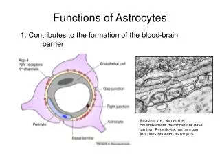
Conduit of ATP Release in Astrocytes B. Corbett Walsh1, George R. Dubyak21Dept. of Physics, Case Western Reserve University, Cleveland, OH 44106 2Dept. of Physiology and Biophysics, Case Western Reserve University School of Medicine, Cleveland, OH 44106 Abstract Methods Discussion Both 1321N1 cells and C6 cells contain thrombin receptors, so when these cells are exposed to thrombin the thrombin is binding to the cell surface. Both cell lines also mobilize Ca2+ as a second messenger in response to being exposed to thrombin. 1321N1 cells clearly contain whatever protein(s) are responsible for causing ATP release when exposed to thrombin because figure 3 and 5 show that they release ATP in response to thrombin. C6 cells however lack the connexin channels sequence in their genome and thus have no way to express it. The C6 cells also do not release ATP when exposed to thrombin as shown in figure 4. These results suggest that connexin channels may be responsible for ATP release when these astrocytes are exposed to thrombin. Cell volume regulation is of particular physiologic importance in neural tissues due to the adverse effects of even small changes in brain volume. In the brain, extracellular ATP acts as an autocrine / paracrine signaling molecule and participates in volume regulation by activating a family of P2 receptors. While significant advances have been made regarding the signaling controlling ATP release, the actual conduits for the export of ATP have not been clearly defined. Investigations of ATP release from astrocytes have identified connexin gap junction hemmichannels as a candidate conduit for ATP release. ATP Assay and transfection using 1321N1 Human Astrocytoma and C6 Rat Glioma are currently underway to determine which conduit is responsible for releasing ATP within astrocytes. A solution is called isotonic if the concentration of osmolytes is the same inside the cell as it is outside. A solution is called hypotonic if more osmolytes are present inside the cell than are present outside of the cell. As a result of this gradient, water will spontaneously diffuse into the cell to correct this imbalance. Because the plasma membrane is a fluid organelle, subject to changes in shape and size, the cell will swell as a result of the sudden intake of water. A solution is called hypertonic if there are more osmolytes outside the cell than inside. Again because of the dynamic nature of the plasma membrane the cell will shrivel up. Luciferase added to Extracellular sample Stimulus, Osmolarity adjustment Rest30 min Isotonic saline Background 10 min Cell monolayer Figure 2. Effects of placing red blood cells in an isotonic solution (left), a hypotonic solution (middle) or hypertonic solution (right). http://biology.unm.edu/ccouncil/Biology_124/Images/tonicity1.jpeg Future Direction Adenosine‐5'‐triphosphate (ATP) is not only a universal energy source constantly produced and utilized by cells, but it also serves as an ‘extracellular second messenger’. ATP, acting as a signaling molecule, participates in many biologic processes. Other functions of ATP include a paracrine role in coordinating hepatocytes and induce apoptosis in various cell types. Since ATP is a relatively large and hydrophilic molecule it thus needs specially designed pathways in order to exit the cells. Experimental 24 well plates were used to simulate conditions under the protocol shown above. Cells were washed of their cell media and allowed to rest for 30 minutes. Then thrombin was added and the correct osmolarity adjustments were given to the appropriate wells. Media surrounding the cells was then extracted and placed in a cuvette with Luciferase (a compound that can be fluorescently detected when bound to ATP). To begin to verify the hypothesis that connexin channels may be responsible for ATP release when C6 and 1321N1 cells are exposed to thrombin under certain osmotic conditions, the C6 cell line will be transfected with the genomic sequence for connexin channels. Figure 6. A basic depiction of transfection; the introduction of DNA into a recipient eukaryotic cell and its subsequent integration into the recipient cells chromosomal DNA. This integration will then allow that DNA sequent to be translated into a protein. http://www.labsupplymall.com/images/products/sg/PolyExpress_Degradation.jpg Once the C6 cells possessed the connexin channels gene, western blotting will be conducted to verify that the C6 cells are expressing that protein on their plasma membrane. Once this has been verified, the same tonicity experiments in the presence and absence of thrombin will be conducted. If ATP is released by the transfected C6 cells, than these connexin channels may be largely responsible for releasing ATP when these cells are exposed to thrombin. Results Plasmid of Connexin Channel to be Introduced to the cell’s genome for expression One possible channel being considered are the connexin gap junction hemmichannels. These channels exist at the point of cell contact and serve as aqueous conduits for ions and hydrophilic molecules smaller than 1kD. Importantly, connexin channels open in response to hypotonic stress and increased intracellular Ca2+, two facts used in the experimental setup. Additionally, numerous studies involving genetic manipulation of connexin subunits have produced evidence that connexin channels can function as ATP release conduits. Expression of Connexin Channel in Cell Genome Figure 1. A rough depiction of a Connexin gap junction Hemmichannel. http://en.wikipedia.org/wiki/ File:Biggapjunct2.png Translation mRNA Figure 4. Experimental results from exposing C6 cells to different osmotic conditions in the presence and absence of thrombin. Figure 3. Experimental results from exposing 1321N1 cells to different osmotic conditions in the presence and absence of thrombin. As shown in figure 3, 1321N1 cells release ATP under hypotonic conditions in the absence of thrombin. Additionally thrombin causes ATP release under isotonic conditions but is inhibited under hypertonic conditions. Hypotonic conditions, when simultaneously exposed to thrombin, potentiate ATP release. Looking at figure 4, C6 cells only release ATP in response to osmotic conditions, specifically hypotonic conditions. This ATP release was not affected at all by the presence of thrombin. Examining the amount of ATP release from both cell lines in figure 5, it becomes evident that the amount of ATP release under similar conditions are comparable to each cell line (comparing C6 +/- thrombin to 1321 – thrombin under any tonicity). Rationale ATP acts as an autocrine / paracrine signaling molecule that has numerous downstream physiological effects. Better understanding of the conduit or pathway of ATP release can enable pharmaceutical companies to develop inhibitors or activators, to enable or prohibit certain processes from occurring. In relation to ATP release in cellular apoptosis, there may be major implications by defining new chemotherapy treatments for cancer patients. It will be crucial to determine which chemotherapies have the capacity to induce immunogenic cell death and which type of tumors can actually generate immunogenic signals after radiotherapy or chemotherapy. Acknowledgments This research was supported in part by NIH grant R01-GM-36387. Hypertonicity Isotonicity Hypotonicity Figure 5. Combining figures 3 (1321N1) and 4(C6) into one graph for easy comparison relative to each other.