Understanding Diffusion MRI: Implications for Tumor Diagnosis and Treatment Monitoring
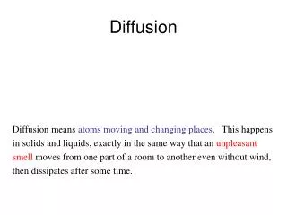
Diffusion MRI (dMRI) is a powerful imaging technique that captures the random motion of water molecules (H2O) in the body, primarily influenced by thermal agitation. This method reveals critical insights into brain tumors by measuring the apparent diffusion coefficient (ADC), which correlates with tumor cell density. High cell density leads to lower ADC values, while low cell density results in higher ADC values. The functional diffusion map (fDM) derived from dMRI is emerging as an early biomarker for response to cytotoxic therapies, highlighting its clinical significance in evaluating tumor progression and treatment efficacy.

Understanding Diffusion MRI: Implications for Tumor Diagnosis and Treatment Monitoring
E N D
Presentation Transcript
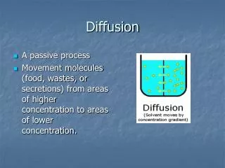
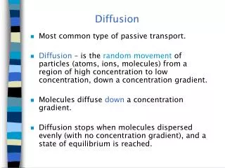
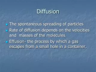
Diffusion Physics • H2O in the body is always in random motion due to thermal agitation B.M. Ellingson, Ph.D., Dept. of Radiological Sciences, David Geffen School of Medicine at UCLA, 2010
Detecting Diffusion with MRI - Intravoxel Incoherent Motion Ellingson et al., Concepts in MR, 2008 From: Ellingson, Concepts in MR, 2008 B.M. Ellingson, Ph.D., Dept. of Radiological Sciences, David Geffen School of Medicine at UCLA, 2010
Detecting Diffusion with MRI - Intravoxel Incoherent Motion Variability in Phase of “Tagged” H2O Level of Diffusion Weighting Detected DWI Signal Diffusion Coefficient MRI Signal w/o Diffusion Sensitivity B.M. Ellingson, Ph.D., Dept. of Radiological Sciences, David Geffen School of Medicine at UCLA, 2010
Image Voxel Proton on H2O = t3 = t2 = t1 MRI Signal Diffusion Time (or level of diffusion weighting) B.M. Ellingson, Ph.D., Dept. of Radiological Sciences, David Geffen School of Medicine at UCLA, 2010
Diffusion MR Characteristics of theCentral Nervous System • Apparent diffusion coefficient (ADC) measured clinically reflects extracellular water • ADC is dependent on tumor cell density (Ellingson, 2010; Sugahara, 1999; Lyng, 2000; Chenevert, 2000; Gaurain, 2001; Nonomura, 2001; Guo, 2002; Chen, 2005; Hayashida, 2006; Manenti, 2008; Kinoshita, 2008 • Cell Density (hypercellular) = ADC • Cell Density (hypocellular) = ADC Edema Necrotic Core Viable Tumor (Dark) ADC Map From: Ellingson, J Magn Reson Imaging, 2010 B.M. Ellingson, Ph.D., Dept. of Radiological Sciences, David Geffen School of Medicine at UCLA, 2010
Diffusion MRI During Successful Cytotoxic Therapy From: Ross, Mol Cancer Ther, 2003 B.M. Ellingson, Ph.D., Dept. of Radiological Sciences, David Geffen School of Medicine at UCLA, 2010
The Functional Diffusion Map (fDM)(Moffat, 2005; 2006; Hamstra, 2005; 2008; Ellingson, 2010) From: Ellingson, JMRI, 2010 B.M. Ellingson, Ph.D., Dept. of Radiological Sciences, David Geffen School of Medicine at UCLA, 2010
Early Detection of Brain Tumor Growth T1+C Contrast-Enhancement (white) FLAIR Hypercellular Regions (Blue) fDMs B.M. Ellingson, Ph.D., Dept. of Radiological Sciences, David Geffen School of Medicine at UCLA, 2010
fDMs in Brain Tumor Progression 3 mo. 6 mo. 9 mo. (Onset of symptoms) T1+C FLAIR fDM B.M. Ellingson, Ph.D., Dept. of Radiological Sciences, David Geffen School of Medicine at UCLA, 2010
fDMs in Progressive Disease (PD) Hypercellularity Hypercellularity Hypercellularity From: Ellingson, ISMRM, 2009; SNO, 2009 B.M. Ellingson, Ph.D., Dept. of Radiological Sciences, David Geffen School of Medicine at UCLA, 2010
Positive Tumor Response to Treatment Day 180 Day 237 Day 298 Day 89 Hypocellular “Treated” Tumor Hypercellular Tumor From: Ellingson, ISMRM, 2009; SNO, 2009 B.M. Ellingson, Ph.D., Dept. of Radiological Sciences, David Geffen School of Medicine at UCLA, 2010
fDM Results in Stable/Responding Disease (SD/RD) Hypocellularity Hypocellularity Hypocellularity From: Ellingson, ISMRM, 2009; SNO, 2009 B.M. Ellingson, Ph.D., Dept. of Radiological Sciences, David Geffen School of Medicine at UCLA, 2010
fDMs May Reflect Molecular/Genetic Phenotypes MGMT(+) MGMT(-) MGMT(+) MGMT(-) MGMT(+) MGMT(-) From: Ellingson, ISMRM, 2009; SNO, 2009 B.M. Ellingson, Ph.D., Dept. of Radiological Sciences, David Geffen School of Medicine at UCLA, 2010
Clinical fDM Sensitivity/Specificity WHO Grade (n = 50 Total Patients) Spearman Corr. Coeff. R = 0.4350, P = 0.0016 B.M. Ellingson, Ph.D., Dept. of Radiological Sciences, David Geffen School of Medicine at UCLA, 2010 From: Ellingson BM et al., ISMRM, 2010
Clinical fDM Sensitivity/Specificity Neurological Status (n = 50 Total Patients) Pearson Corr. Coeff. R2 = 0.8586, P < 0.0001 B.M. Ellingson, Ph.D., Dept. of Radiological Sciences, David Geffen School of Medicine at UCLA, 2010 From: Ellingson BM et al., ISMRM, 2010
fDMs as an early biomarker for cytotoxic and new anti-angiogenic treatments B.M. Ellingson, Ph.D., Dept. of Radiological Sciences, David Geffen School of Medicine at UCLA, 2010 From: Ellingson BM, J Neuroonc, 2010
Volumetric Analysis of fDMs as an early biomarker for cytotoxic and new anti-angiogenic treatments Bevacizumab fDMs detect PD > 2 months before recurrence Temozolomide fDMs predict survival and progression better than age and tumor grade! B.M. Ellingson, Ph.D., Dept. of Radiological Sciences, David Geffen School of Medicine at UCLA, 2010 From: Ellingson BM, J Neuroonc, 2010
Better Defining ADC Thresholds for Classification ADC = 95% C.I. NAWM+NAGM ADC = 95% C.I. NAWM ADC = 95% C.I. NAWM+NAGM+CSF ADC = 95% C.I. NAGM Different ADC thresholds reflect different sensitivity/specificity to growing tumor From: Ellingson, JMRI, 2010 B.M. Ellingson, Ph.D., Dept. of Radiological Sciences, David Geffen School of Medicine at UCLA, 2010
Better Defining ADC Thresholds for Classification Different ADC thresholds reflect different sensitivity/specificity to growing tumor From: Ellingson, JMRI, 2010 B.M. Ellingson, Ph.D., Dept. of Radiological Sciences, David Geffen School of Medicine at UCLA, 2010
Graded fDMs Allow Visualization and Quantification of Growing Tumor + Hypocellular + Hypercellular Biological Calibration From: Ellingson, JMRI, 2010 B.M. Ellingson, Ph.D., Dept. of Radiological Sciences, David Geffen School of Medicine at UCLA, 2010
Graded fDMs Allow Better Visualization of Growing Tumor + Hypocellular + Hypercellular B.M. Ellingson, Ph.D., Dept. of Radiological Sciences, David Geffen School of Medicine at UCLA, 2010
Graded fDMs Allow Better Visualization of Growing Tumor B.M. Ellingson, Ph.D., Dept. of Radiological Sciences, David Geffen School of Medicine at UCLA, 2010
Graded fDMs in Differential DiagnosisTumor vs. Demyelination Hypercellular Hypocellular Macrophages & Inflammatory Cells Demyelination Biopsy Diagnosis = Demyelination (Multiple Sclerosis) B.M. Ellingson, Ph.D., Dept. of Radiological Sciences, David Geffen School of Medicine at UCLA, 2010
Graded fDMs can distinguish radiation necrosis from tumor T1+C FLAIR Hypercellular Graded fDM Hypocellular B.M. Ellingson, Ph.D., Dept. of Radiological Sciences, David Geffen School of Medicine at UCLA, 2010
Diffusivity Mismatch Index (DMI) predicts survival From: Ellingson, Clin Cancer Res, 2010, Submitted B.M. Ellingson, Ph.D., Dept. of Radiological Sciences, David Geffen School of Medicine at UCLA, 2010
Diffusivity Mismatch Index (DMI) predicts survival From: Ellingson, Clin Cancer Res, 2010, Submitted B.M. Ellingson, Ph.D., Dept. of Radiological Sciences, David Geffen School of Medicine at UCLA, 2010
Cell Invasion, Migration, and Proliferation Level Estimates = CIMPLE Maps • Higher-order changes in ADC over time and space • Allows us to map estimates of invasion and proliferation From: Ellingson, Magn Reson Med, 2010, Accepted B.M. Ellingson, Ph.D., Dept. of Radiological Sciences, David Geffen School of Medicine at UCLA, 2010
Whole Brain CIMPLE Maps & 18F-FDOPA PET From: Ellingson, Magn Reson Med, 2010, Accepted B.M. Ellingson, Ph.D., Dept. of Radiological Sciences, David Geffen School of Medicine at UCLA, 2010
Whole Brain CIMPLE Maps & 18F-FDOPA PET -10 10 Cell Proliferation [1/yr] B.M. Ellingson, Ph.D., Dept. of Radiological Sciences, David Geffen School of Medicine at UCLA, 2010
CIMPLE Maps Post-Tx Recurrence Pre-Tx From: Ellingson, Cancer Imaging, 2010, Submitted B.M. Ellingson, Ph.D., Dept. of Radiological Sciences, David Geffen School of Medicine at UCLA, 2010
CIMPLE Maps (Doubling Times) • Within physiologic range of doubling times: • 22 days (GBM) - 556 days (WHO II) Blankenberg et al, AJNR, 2005 Doubling Time Days 0 365 B.M. Ellingson, Ph.D., Dept. of Radiological Sciences, David Geffen School of Medicine at UCLA, 2010
Doubling Time CIMPLE Maps (Doubling Times) Days 0 365 Doubling Time (Days) < 10 20 30 40 50 B.M. Ellingson, Ph.D., Dept. of Radiological Sciences, David Geffen School of Medicine at UCLA, 2010
Mean proliferation at start of treatment predicts survival (defined from time of CIMPLE map) N = 26 From: Ellingson, Cancer Imaging, 2010, Submitted B.M. Ellingson, Ph.D., Dept. of Radiological Sciences, David Geffen School of Medicine at UCLA, 2010
Diffusion MRI is sensitive to cell density Functional diffusion maps (fDMs) reflect voxel-by-voxel changes in cellularity fDM kinetics are useful for individual patient monitoring Conclusions B.M. Ellingson, Ph.D., Dept. of Radiological Sciences, David Geffen School of Medicine at UCLA, 2010
Graded fDMs allow for degree of cellularity to be visualized and quantified (biological basis) Graded fDMs can distinguish radiation necrosis from tumor recurrence CIMPLE maps allow visualization and quantification of invasion and proliferation rates Conclusions B.M. Ellingson, Ph.D., Dept. of Radiological Sciences, David Geffen School of Medicine at UCLA, 2010
Radiology Whitney Pope, M.D., Ph.D. Dieter Enzmann, M.D. Jonathan Goldin, M.D. MedQIA Neurology/Neuro-Oncology Timothy Cloughesy, M.D. Albert Lai, M.D., Ph.D. Neurosurgery Linda Liau, M.D. Bob Shafa, M.D. Antonio DeSalles, M.D. Pathology Paul Mischel, M.D. Bill Yong, M.D. Thank You! Radiology • Kathleen Schmainda, Ph.D. • Scott Rand, M.D., Ph.D. Neurology/Neuro-Oncology • Mark Malkin, M.D. • Jennifer Connelly, M.D. Neurosurgery • Wade Mueller, M.D. • Shekar Kurpad, M.D., Ph.D. B.M. Ellingson, Ph.D., Dept. of Radiological Sciences, David Geffen School of Medicine at UCLA, 2010