Download
1 / 33
380 likes | 1.29k Views
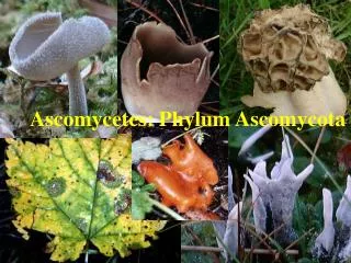
Filamentous Ascomycetes Eurotiales and Allied Taxa. Pl P 421/521 Lecture 6. From Spatafora et al. 2006. A five-gene phylogeny of Pezizomycotina. Mycologia 98: 1018-1028. From Geiser et al. 2006. Mycologia 98: 1063-1064. Onygenales. Family Onygenaceae

E N D
Filamentous AscomycetesEurotiales and Allied Taxa Pl P 421/521 Lecture 6
From Spatafora et al. 2006. A five-gene phylogeny of Pezizomycotina. Mycologia 98: 1018-1028
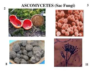
Onygenales • Family Onygenaceae • Onygena equina on old hooves and horns • Stalked ascomata called mazaedia • Ajellomyces dermatidis—blastomycosis and Ajellomyces capsulatus—histoplasmosis • Dimorphic, growing as yeasts at 37C and mycelium below 37C • Coccidioidesimmitis —Valley Fever (Coccidioidomycosis) • Produces arthrospores in soil, and enlarged, multinucleate spherules in host tissue
Onygena equina Stalked ascomata (mazaedia)
Coccidiodes immitis Arthrospores (above), sphaerules formed in host tissue (right)
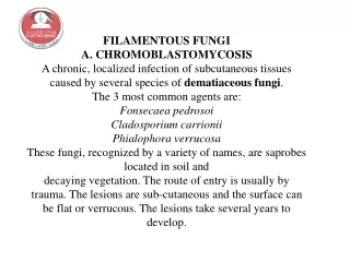
Arthrodermataceae • Arthroderma—cleistothecia with bone-shaped cells in walls (ossiform cells) • Trichophyton and Microsporum anamorphs • Dermatophytes—fungi that grow in the dead layer of keratinized skin • Ringworm • Athlete’s foot • Nail fungus
Eurotiales (Plectomycetes) • Thin-walled asci (prototunicate) • Asci scattered within cleistothecium • One-celled ascospores • Two familes: • Trichocomaceae • Aspergillus, Penicillium and Paecilomyces anamorphs • Pseudoeurotiaceae
Aspergillus Conidia Conidiogenous cells (phialides) Supporting cell (branch or metulae) Swollen apex of conidiophore (vesicle) Conidiophore Basal part of conidiophore (foot cell)
Anamorphs--Aspergillus SEM by Charles Mims
Genus/Species:Aspergillus flavus • Image Type:Microscopic Morphology • Title:Stages in development of fruiting bodies • Disease(s): Aspergillosis • Legend:Stages in development of fruiting bodies. Differential interference contrast microscopy, 630X. Aspergillus flavus http://www.doctorfungus.org
Anamorphs--Penicillium phialides Branches (metulae)
Anamorphs--Paecilomyces Divergent phialides with swollen base and long, tapering neck Colonies may be pink, purple, yellow, brown or white, but never green as in Penicillium spp.
Teleomorphs • Aspergillus: • Eurotium • Neosartorya • Emericella • Penicillium: • Eupenicillium • Talaromyces • Paecilomyces: • Byssochlamys
Eurotium • Aspergillus anamorph • Cleistothecia yellow to orange-red • wall composed of single layer of flattened cells • ascospores flattened, usually with equatorial groove. Ascospore by D. Geiser From Hanlin, 1998. Illustrated Genera of Ascomycetes Vol II
Emericella • Aspergillus anamorph • Cleistothecial wall surrounded by hülle cells • Ascospores small, colored, lens-shaped with flange From Hanlin, 1998. Illustrated Genera of Ascomycetes Vol II
Emericella Hülle cells, D. Geiser
Eupenicillium • Penicillium anamorph • Cleisothecia hard, white becoming colored (yellow, orange, brown) • Ascospores small, hyaline or yellowish, lens-shaped, often with equatorial flanges From Hanlin, 1998. Illustrated Genera of Ascomycetes Vol II
Talaromyces • Paecilomyces or Penicillium anamorph • Cleistothecium whitish to bright yellow • Wall composed of interwoven hyphae • Ascospores ellipsoidal, with spiny walls From Hanlin, 1998. Illustrated Genera of Ascomycetes Vol II
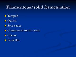
The good and the bad • Penicillium spp.—antibiotic production • Penicilliumroqueforti—blue cheese • Penicillium spp.—blue and green molds on bread, cheese, fruits, vegetables • Aspergillus flavus—aflatoxins (moldy peanuts) • A. flavus/A. niger--aspergillosis
Penicillin • Penicillium notatum growing in Alexander Fleming’s Petri dish of Staphylococcus in 1928 led to the discovery of penicillin • Howard Florey & Ernest Chain (1939) began work on purification and trials • 1941—work moved to US (NRRL in Peoria, IL) to escape bombing in London (WWII) • Fermentation vessels and corn steep liquor • Mary Hunt (“Moldy Mary”) brought in P. chyrogenum on a melon • 1945—Fleming, Florey & Chain received Noble Prize
Penicillium notatum Penicillin prevents cross-linking of small peptide chains in peptidoglycan, the main wall polymer in bacteria. Newly formed cells are abnormal in shape and susceptible to osmotic lysis.