Download
1 / 2
20 likes | 36 Views
Mammography / Tomosynthesisis the recommended first step in breast cancer screening for all women aged 40 years and older except those who are pregnant. Mammogram helps to detect 2 to 7 cancers in women’s breasts.

E N D
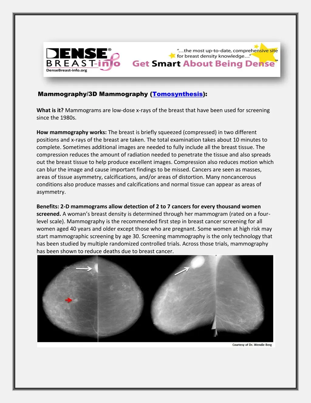
Mammography/3D Mammography (Tomosynthesis): What is it? Mammograms are low-dose x-rays of the breast that have been used for screening since the 1980s. How mammography works: The breast is briefly squeezed (compressed) in two different positions and x-rays of the breast are taken. The total examination takes about 10 minutes to complete. Sometimes additional images are needed to fully include all the breast tissue. The compression reduces the amount of radiation needed to penetrate the tissue and also spreads out the breast tissue to help produce excellent images. Compression also reduces motion which can blur the image and cause important findings to be missed. Cancers are seen as masses, areas of tissue asymmetry, calcifications, and/or areas of distortion. Many noncancerous conditions also produce masses and calcifications and normal tissue can appear as areas of asymmetry. Benefits: 2-D mammograms allow detection of 2 to 7 cancers for every thousand women screened. A woman’s breast density is determined through her mammogram (rated on a four- level scale). Mammography is the recommended first step in breast cancer screening for all women aged 40 years and older except those who are pregnant. Some women at high risk may start mammographic screening by age 30. Screening mammography is the only technology that has been studied by multiple randomized controlled trials. Across those trials, mammography has been shown to reduce deaths due to breast cancer.
Figure 1. Analog (Film) Mammograms from a 62-year-old female with lump felt under her right arm. The breasts are not dense, with only scattered fibroglandular density. Left-hand images show views from above (known as craniocaudal or “CC” views) and right-hand images are taken from a side angle (known as mediolateral oblique or “MLO” views) and show a dense mass in right underarm (axilla, white arrow, triangle marker). Ultrasound-guided biopsy showed this mass to be a lymph node involved with cancer spread from the breast (i.e. a metastatic lymph node). The primary cancer in the right breast itself was not initially seen on these images but can be seen in retrospect on the CC view only (short red arrow). 2) Digital, 2-Dimensional, known as “Full Field Digital Mammogram” (FFDM), which uses a dedicated electronic detector system to computerize and display the x-ray information (Fig. 2). Figure 2. Digital CC and MLO Mammograms from the same patient as in Figure 1 again show the metastatic cancerous lymph node (arrows). Better seen is a subtle mass with associated distortion (red ovals) in the upper inner right breast. The skin and tissues near the skin are also better seen on digital mammography than on film. 3) Tomosynthesis, also referred to as “3-Dimensional mammography” (3D mammography) or “tomo”, uses a dedicated electronic detector system to obtain multiple projection images which are “synthesized” by the computer to create thin slices of the breast. Contact As DenseBreast-info, Inc. PO Box 997 Deer Park, NY 11729 http://densebreast-info.org