Download
1 / 1
10 likes | 115 Views
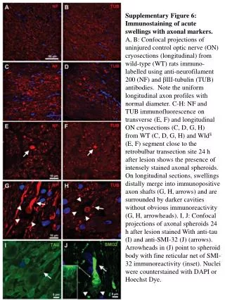
Supplementary Figure 6: Immunostaining of acute swellings with axonal markers. A, B: Confocal projections of uninjured control optic nerve (ON) cryosections (longitudinal) from wild-type (WT) rats immuno - labelled using anti-neurofilament 200 (NF) and β III - tubulin (TUB)

E N D
Supplementary Figure 6: Immunostaining of acute swellings with axonal markers. A, B: Confocal projections of uninjured control optic nerve (ON) cryosections (longitudinal) from wild-type (WT) rats immuno- labelled using anti-neurofilament 200 (NF) and βIII-tubulin (TUB) antibodies. Note the uniform longitudinal axon profiles with normal diameter. C-H: NF and TUB immunofluorescence on transverse (E, F) and longitudinal ON cryosections (C, D, G, H) from WT (C, D, G, H) and WldS (E, F) segment close to the retrobulbar transection site 24 h after lesion shows the presence of intensely stained axonal spheroids. On longitudinal sections, swellings distally merge into immunopositive axon shafts (G, H, arrows) and are surrounded by darker cavities without obvious immunoreactivity (G, H, arrowheads). I, J: Confocal projections of axonal spheroids 24 h after lesion stained With anti-tau (I) and anti-SMI-32 (J) (arrows). Arrowheads in (J) point to spheroid body with fine reticular net of SMI-32 immunoreactivity (inset). Nuclei were counterstained with DAPI or Hoechst Dye.