Download
1 / 39
440 likes | 1.18k Views
Acute Mesenteric Ischemia and Infarction. foolad Eghbali M.D. Vascular surgeon Rasool Akram Hosp. Background. Acute mesenteric ischemia (AMI) is a syndrome in which inadequate blood flow through the mesenteric vessels causes ischemia and eventual gangrene of the bowel wall.

E N D
Acute Mesenteric Ischemia and Infarction fooladEghbali M.D. Vascular surgeon RasoolAkram Hosp.
Background • Acute mesenteric ischemia (AMI) is a syndrome in which inadequate blood flow through the mesenteric vessels causes ischemia and eventual gangrene of the bowel wall. • Either arterial or venous disease
Arterial disease may be subdivided into nonocclusive mesenteric ischemia (NOMI)and occlusive mesenteric arterial ischemia (OMAI). • OMAI may be further subdivided into acute mesenteric arterial embolus (AMAE) and acute mesenteric arterial thrombosis (AMAT). Venous disease takes the form of mesenteric venous thrombosis (MVT). • AMI comprises 4 different primary clinical entities: NOMI, AMAE, AMAT, and MVT.
since 1930, many advances have been made that allow earlier diagnosis and treatment. • Whereas the prognosis remains grave
Superior Mesenteric Artery (SMA) Largest caliber vessel + 45-degree angle makes it most commonly occluded Aorta Celiac Trunk SMA IMA
The celiac artery (CA) supplies the foregut, hepatobiliary system, and spleen; the SMA supplies the midgut (ie, small intestine and proximal mid colon); and the inferior mesenteric artery (IMA) supplies the hindgut (ie, distal colon and rectum).However, multiple anatomic variants are observed. Venous drainage is through the superior mesenteric vein (SMV), which joins the splenic vein to form the portal vein
Superior Mesenteric Artery (SMA) • Emboli occlude past the middle colic, causing small bowel ischemia Middle Colic SMA Jejunal & Ileal Arteries Occlusion Point Right Colic Ileocolic
Pathophysiology • Insufficient perfusion of the small bowel and colon may result from arterial occlusion by embolus or thrombosis (AMAE or AMAT), thrombosis of the venous system (MVT), or nonocclusive processes such as vasospasm or low cardiac output (NOMI).
Etiologies of Acute Mesenteric Ischemia (AMI) • SMA Occlusion (at least 60% of cases) • Embolism: MI, Afib, Endocarditis, Valve d. • Thrombosis: Atherosclerosis – plaque rupture • Nonocclusive Mesenteric Ischemia (NOMI) • Atherosclerosis + shock + vasopressors • Mesenteric Venous Thrombosis (MVT) • Primary clotting disorder
Etiologies of Acute Mesenteric Ischemia (AMI) • Focal small bowel ischemia - rare • Partial malrotation, volvulus, mesenteric hematoma, strangulated hernia • Unknown • ?Mesenteric small vessel disease
Causes of embolic AMI (AMAE) include the following: • Cardiac emboli - Mural thrombus after myocardial infarction, auricular thrombus associated with mitral stenosis and atrial fibrillation, septic emboli from valvularendocarditis (less frequent) • Emboli from fragments of proximal aortic thrombus due to a ruptured atheromatous plaque • Atheromatous plaque dislodged by arterial catheterization
Causes of thrombotic AMI (AMAT) include the following: • Atherosclerotic vascular disease (most common) • Aortic aneurysm • Aortic dissection • Arteritis • Decreased cardiac output from myocardial infarction or CHF (thrombotic AMI may cause acute decompensation) • Dehydration from other causes
Causes of NOMI include the following: • Hypotension from CHF, myocardial infarction, sepsis, aortic insufficiency, severe liver or renal disease, or recent major cardiac or abdominal surgery • Vasopressive drugs • Ergotamines • Cocaine • Digitalis (whether digitalis use causes NOMI or patients who develop NOMI are older and are more likely to have been prescribed digitalis is unclear)
Causes of MVT include the following (>80% of patients with MVT are found to have predisposing conditions): • Hypercoagulability from protein C and S deficiency, antithrombin III deficiency, dysfibrinogenemia, abnormal plasminogen, polycythemiavera (most common), thrombocytosis, sickle cell disease, factor V Leiden mutation, pregnancy, and oral contraceptive use • Tumor causing venous compression or hypercoagulability (paraneoplastic syndrome) • Infection, usually intra-abdominal (eg, appendicitis, diverticulitis, or abscess) • Venous congestion from cirrhosis (portal hypertension) • Venous trauma from accidents or surgery, especially portocaval surgery • Increased intra-abdominal pressure from pneumoperitoneum during laparoscopic surgery • Pancreatitis
Epidemiology • Age • Advanced age is a risk factor due to the association with atheroscleosis • The overall prevalence of AMI is 0.1% of all hospital admissions • No overall sex preference
Prognosis • The prognosis of AMI of any type is grave. Overall, the mortality rate in the last 15 years from all causes of AMI averages 71%, with a range of 59-93%. Once bowel wall infarction has occurred, the mortality rate is as high as 90%. Even with good treatment, up to 50-80% of patients die.
History & Physical Classic Presentation: • Rapid onset of severe, unrelenting periumbilical pain • Pain out of proportion to findings on physical examination. • Nausea and vomiting • Forceful/urgent bowel evacuation • Risk factors for acute mesenteric ischemia
History & Physical SMA Thrombosis: • Prodrome of postprandial pain/nausea and weight loss • Presentation with classic symptoms Non-occlusive Mesenteric Ischemia: • Unexplained decline in clinical status or failure to follow expected recovery
History & Physical Mesenteric Venous Thrombosis: • Fever • Abdominal distension • Hemoccult positive stool
Physical Examination • The different etiologies notwithstanding, physical examination findings are generally similar in patients with AMI. The main distinction is between early and late presentation. Early in the course of the disease, in the absence of peritonitis, physical signs are few and nonspecific. Tenderness is minimal to nonexistent. Stool may be guaiac positive.
Peritoneal signs develop late, when infarction with necrosis or perforation occurs. Tenderness becomes severe and may indicate the location of the infarcted bowel segment. A palpable tender mass may be present. Bowel sounds range from hyperactive to absent. Voluntary and involuntary guarding appears. Fever, hypotension, tachycardia, tachypnea, and altered mental status are observed. Foul breath may be noted with bowel infarction, from the putrefaction of undigested alimentary material accumulated proximal to the pathologic site • Signs reflecting risk factors for AMI may be noted.
Complications • Bowel necrosis necessitating bowel resection • Septic shock • Death
Diagnostic Considerations • Because acute mesenteric ischemia (AMI) is a condition with an unclear initial presentation, serious morbidity, and a high mortality rate without proper treatment, clinical suspicion should remain high. Obtain early angiography if any suspicion of AMI exists. Subsequent treatment should be initiated as rapidly as possible. No patient in whom AMI is suspected should be discharged unless AMI can be ruled out.
Laboratory Findings • Anion gap metabolic acidosis • Elevated arterial/venous lactate • Leukocytosis • Hemoconcentration • Elevated LDH, amylase, AST, and CPK • Elevated K and Phos are late signs
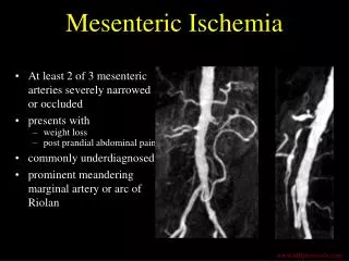
Radiology • Plain films – thumbprinting, thickened bowel (<40% sensitivity) • CT – thickened/dilated bowel, intramural hematoma, pneumatosis (64% sensitivity) • MRI – promising but untested to date • Mesenteric angiography – test of choice; can identify the type of AMI
Differential Diagnosis Other serious conditions to consider: • Pancreatitis • Acute Diverticulitis • Acute Cholecystitis • Small bowel obstruction • Perforation of a viscous • Ruptured aneurysm
Treatment • Resuscitation with fluids/blood products • Anticoagulation, Administer heparin as a bolus of 80 U/kg, and then as an infusion at 18 U/kg/h until full conversion to oral warfarin • Infusion of a vasodilator • Glucagon systemically OR • Papaverine through a catheter, Start an infusion of 30-60 mg/h after angiography,
Inpatient medications include the following: • Papaverine - For patients with arterial occlusive AMI or nonocclusive mesenteric ischemia (NOMI) • Heparin - For patients who have mesenteric venous thrombosis (MVT) or have undergone revascularization • Warfarin - For long-term treatment of patients with MVT or atrial fibrillation • Broad-spectrum antibiotics and pain medications - For all patients • Thrombolytics - For selected patients with embolic AMI • Some experience with percutaneous endovascular interventions has been accumulated. In select cases, especially in isolated spontaneous dissection of the SMA, stent placement may offer the best option
Surgical Care • Before operative management of AMI, stabilize patients by means of intravenous (IV) fluid administration, antibiotic prophylaxis covering the colonic flora, nasogastric tube decompression, and bladder catheterization, with heparin or papaverine administered as indicated. Blood should be available
In all types of AMI, resection of necrotic bowel may be required if signs of peritonitis develop. Differentiation of nonviable from viable bowel can be enhanced by intraoperativefluorescein administration