Download
1 / 33
330 likes | 493 Views
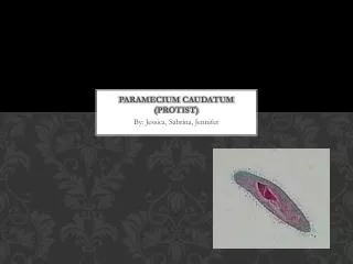
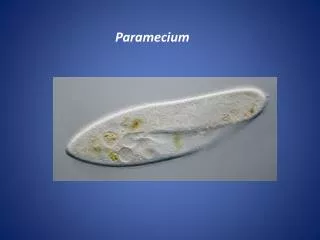
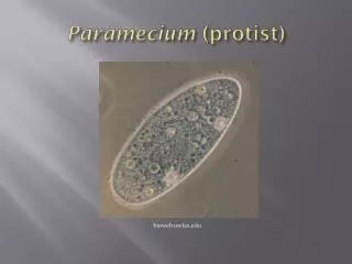
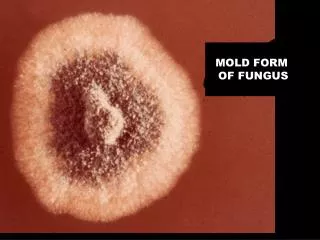
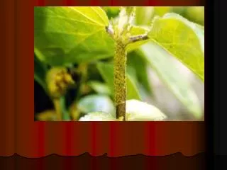
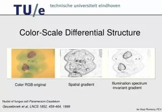
Color-Scale Differential Structure. Illumination spectrum -invariant gradient. Spatial gradient. Color RGB original. Nuclei of fungus cell Paramecium Caudatum. Geusebroek et al, LNCS 1852, 459-464, 1999. The color of an object depends on color of the illuminating light

E N D
Color-Scale Differential Structure Illumination spectrum -invariant gradient Spatial gradient Color RGB original Nuclei of fungus cell Paramecium Caudatum Geusebroek et al, LNCS 1852, 459-464, 1999
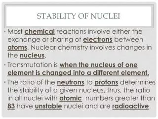
The color of an object depends on • color of the illuminating light • illumination intensity • sensor sensitivity • direction of surface normal • surface reflectance properties • Assumptions: • Scene is uniformly illuminated • light source is colored • surface has Lambertian reflectance
What causes color ? object color spectral color Lamp
Emission spectrum of black body radiator Spectrum reflected froman arbitrary object Object reflectance function for the observed spectrum for a resp. 2500K, 6500K and 10,000K light source:
0 1 2 Color receptive fields 0 1 2
Self-organization: receptive fields from Eigenpatches(12x12 pixels)
0 1 2 Colour receptivefields fromEigenpatches
0.3 0.2 0.1 0 -0.1 -0.2 -0.3 -0.4 0 10 20 30 40 50 60 70 80 90 Hering basis How can we measure color? RF sensitivity wavelength Idea Koenderink: Gaussian derivatives of zero, first and second order in the wavelength domain
0.9 0.8 L 0.7 0.6 M 0.5 0.4 0.3 S 0.2 0.1 0 300 400 500 600 700 800 900 Taylor color model Cone sensitivity Luminance Blue-yellowness Purple-greenness
s Spatial color Energy densities cannot be measured at a point, … … one probes a certain volume Color scale-space starts by probing this space.
Reflectance of light What are invariant properties? object color spectral color Lamp
The reflected spectrum is: v = viewing direction n = surface patch normal s = direction of illumination f = Fresnel front surface reflectance coefficient in v R = body reflectance
Because of projection of the energy distribution on the image plane the vectors n, s and v will depend on the position at the imaging plane. So the energy at a point x is then related to: We assume an illumination with a locally constant color:
Aim: describe material changes independent of the illumination. Bothequationshavemanycommonterms
The normalized differential determines material changes independent of the viewpoint, surface orientation, illumination direction, illumination intensity and illumination color!
The derivative jet to x and forms a complete family of geometric invariants: These are observed properties, so we convolve with Gaussian derivatives
Color invariants Color edges can be defined as the thresholding of the spatial gradient (color-invariant equivalent of Lw):
Spatial color model and tracing color edges in microscopy Influence of illumination color temperature on edge strength, scale is 3.0 px. Skin tissue section illuminated by a halogen bulb at 4000 K (top) and 2600 K (bottom) color temperature.
Some color differential invariants
Feulgen stain,red-green edges Paramecium caudatum, Feulgen and Fast green stain Color canny, red-green normalized edges, scale 3
Hematoxylin eosin stain Pituitary gland, sheep, adenohypophysis 40x Cell: E<0, E > 0, scale 1.0 Nuclei: E <0, E > 0, E +E < 0, scale 3.0 additional constraint added to refine selection
Safranin O stain E > 0, E > 0, scale sigma 1.0 Safranin O stain for proteoglycans (mouse knee joint) Courtesy of Koen Gijbels and Paul Stoppie
Oil red O stain Oil red O stain of fat emboli in lung E > 0, E > 0, scale 1.5
PAS stain Lww > 0, Lvv Lww-Lvw2 > 0, E -E > 0, scale sigma 2.0 P.A.S. stain for carbohydrates (goblet cells, gut) carbohydrates stain magenta - elliptic patches
Blood smear Blood smear, Giemsa stain, 100x, JPEG compression RBC: E > 0, E +E > 0, scale 0.5 Leucocytes: E < 0, scale 12 Leucocyte nuclei: E < 0, E > 0, scale 3
Blue-yellow edges Note the complete absence of detection of black-white edges.
Second order color invariants Color edges can also be defined as the zero-crossings of the second order derivative in the spatial gradient direction (color-invariant equivalent of Lww):
Color invariant edge detection Luminance gradient edge detection
Conclusions • Color ‘scale-space’ compatible with classical luminance scale-space • The model enables the design of practical image analysis ‘color reasoning’ solutions, e.g. invariance for illumination • The color-scale invariant differential operators are building blocks for a differential geometry on color images