Download
1 / 46
490 likes | 1.02k Views
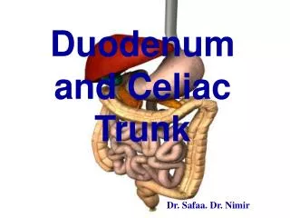
Duodenum and Celiac Trunk Dr. Safaa. Dr. Nimir. Objectives. Describe the surface anatomy of the duodenum. Enumerate parts of the duodenum. Discuss anatomical relations of the duodenum. What structures open in the second part of the duodenum. Explain the blood supply of the duodenum.

E N D
Duodenum and Celiac Trunk Dr. Safaa. Dr. Nimir
Objectives • Describe the surface anatomy of the duodenum. • Enumerate parts of the duodenum. • Discuss anatomical relations of the duodenum. • What structures open in the second part of the duodenum. • Explain the blood supply of the duodenum. • Define celiac trunk. • Enumerate the branches of celiac trunk.
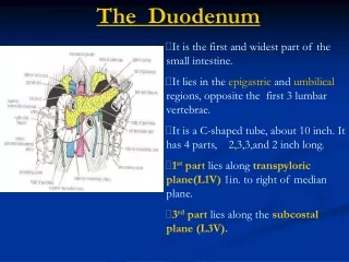
Duodenum Dr W@el
Parts 1st part 2nd part 4th part 3rd part
Length 2 inch 1 inch 3 inch 4 inch
Superior (first) part: • Relations • 2. The second inch of the duodenum: • • Anteriorly: • The quadrate lobe of the liver, • The neck of the gall bladder • • Posteriorly: • a. The bile duct. • b. The gastroduodenal artery. • c. The portal vein. • d. The inferior vena cava. • • Superiorly: the opening into the lesser sac. • • Inferiorly: pancreas.
Superior (first) part: • Relations • 2. The second inch of the duodenum: • • Anteriorly: • The quadrate lobe of the liver, • The neck of the gall bladder • • Posteriorly: • a. The bile duct. • b. The gastroduodenal artery. • c. The portal vein. • d. The inferior vena csva. • • Superiorly: the opening into the lesser sac. • • Inferiorly: pancreas.
Superior (first) part: • Relations • 2. The second inch of the duodenum: • • Anteriorly: • The quadrate lobe of the liver, • The neck of the gall bladder • • Posteriorly: • a. The bile duct. • b. The gastroduodenal artery. • c. The portal vein. • d. The inferior vena csva. • • Superiorly: the opening into the lesser sac. • • Inferiorly: pancreas.
Second part: Length: three inches long. Extent: from the neck of gall bladder to L3 Direction: descends vertically. Peritoneal covering: covered by the peritoneum only on the anterior surface except the middle part where is crossed by transverse colon.
Second part: • Relations of the second part: • • Anteriorly: • Upper part: right lobe of the liver. • The middle part: transverse colon. • Lower part: loops of the jejunum. • • Laterally: • The right colic flexure. • The fat in front of the right kidney. • • Medially: • The head of pancreas • The bile duct • • Posteriorly: • The hilum of right kidney. • The right renal vessels. • The right psoas major muscle. • .
Second part: • Relations of the second part: • • Anteriorly: • Upper part: right lobe of the liver. • The middle part: transverse colon. • Lower part: loops of the jejunum. • • Laterally: • The right colic flexure. • The fat in front of the right kidney. • •Medially: • The head of pancreas • The bile duct • • Posteriorly: • The hilum of right kidney. • The right renal vessels. • The right psoas major muscle. • .
Third part: Length: four inches Extent: at the level of the 3rd lumbar vertebra Direction: horizontal. Peritoneal covering: is only covered by peritoneum anteriorly and inferiorly except the site of attachment of mesentry.
Third part: Length: four inches Extent: at the level of the 3rd lumbar vertebra. Direction: lies in a horizontal plane, Peritoneal covering: is only covered by peritoneum anteriorly and inferiorly except the site of attachment of mesentry.
Third part: • Relations of the 3rd part: • • Anteriorly: • The root of the mesentery. • The coils of jejunum. • • Posteriorly: • The right ureter. • The right psoas major muscle. • The right testicular (or ovarian) vessels. • Inferior vena cava. • Abdominal aorta & origin of the inferior mesenteric artery. • • Superiorly: • The pancrcas. • • Inferiorly: • Coils of the jejunum.
Fourth part: Length: one inch long. Extent: from the level of the 3rd to the level of the 2nd lumbar vertebrae. Direction: ascends to end by forming the duodenojejunal flexure. Peritoneal covering: is covered by the peritoneum anteriorly and to the left.
Fourth part: Length: one inch long. Extent: from the leve] of the 3rd to the level of the 2nd lumbar vertebrae. Direction: ascends to end by forming the duodenojejunal flexure. Peritoneal covering: is covered by the peritoneum anteriorly and to the left.
Fourth part: Length: one inch long. Extent: from the leve] of the 3rd to the level of the 2nd lumbar vertebrae. Direction: ascends to end by forming the duodenojejunal flexure. Peritoneal covering: is covered by the peritoneum anteriorly and to the left.
Structures opening in the 2nd part of the duodenum • The bile duct unites with the pancreatic duct forming a dilatation called the hepatopancreatic ampulla (ampulla of Vater). • The ampulla opens on an elevation called the major duodenal papilla. • The accessory pancreatic duct opens one-inch above the major duodenal papilla, forming a smaller elevation called the minor duodenal papilla.
Structures opening in the 2nd part of the duodenum • The bile duct unites with the pancreatic duct forming a dilatation called the hepatopancreatic ampulla (ampulla of Vater). • The ampulla opens on an elevation called the major duodenal papilla. • The accessory pancreatic duct opens one-inch above the major duodenal papilla, forming a smaller elevation called the minor duodenal papilla.
The primitive gut tube can be divided into foregut, midgut, and hindgut regions. • The foregut is supplied by the celiac trunk. • The midgut is supplied by the superior mesenteric artery . • The hindgut is supplied by the inferior mesenteric artery .
The three anterior branches supply the gastrointestinal viscera: • the celiac trunk • superior mesenteric artey • inferior mesenteric artery
The celiac trunk Arises from the abdominal aorta anterior to the upper part of vertebra LI. It divides into: Gastric Splenic Common hepatic arteries
Theleft gastric artery : • The left gastric artery supplies the lesser curvature of the stomach and anastomoses with the right gastric artery. • eophageal branches (anastomose with esophageal branches from the thoracic aorta).
The splenic artery takes a tortuous course to the left along the superior border of the pancreas . • The splenic artery gives off • short gastric arteries • left gastro-epiploic artery
The common hepatic artery divides into its two terminal branches, • the hepatic artery proper and divides into the i.right hepatic artery ii.left hepatic artery • gastroduodenal artery: • right gastro-epiploic(omental )artery • superior pancreaticoduodenal artery
Blood supply of the duodenum • The superior pancreaticoduodenal artery arises from the gastroduodenal artery • inferior pacreaticoduodenal artery originates as the first branch of the superior mesenteric artery. • These vessels both lie in the curve between the duodenum and the head of the pancreas,
THANK YOU Dr W@el