Microscopy Techniques and Illumination Methods: A Comprehensive Overview
Explore the world of microscopy with details on objective and eyepiece magnification, illumination techniques like bright field and Köhler, staining methods, and phase contrast microscopy. Learn about enhancing contrast and optimizing illumination for better microscopy results.

Microscopy Techniques and Illumination Methods: A Comprehensive Overview
E N D
Presentation Transcript
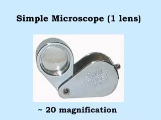
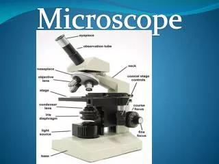
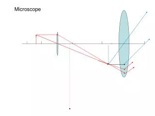
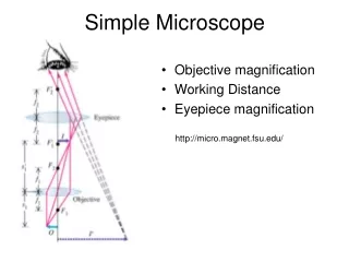
Simple Microscope • Objective magnification • Working Distance • Eyepiece magnification http://micro.magnet.fsu.edu/
Illumination (Bright Field) Summary the field of view should be (reasonably) evenly illuminated the illuminating train should be able to fully illuminate the aperture of an objective of NA = 1.0 the light source should be focused in the object in critical illumination, the light bulb is the light source in Köhler illumination, the light source is an iris diaphragm attached to the illuminator (the field stop) the condenser iris is adjusted for each objective • Simple mirror (historical microscope) • Critical illumination • Koehler Illumination
Staining • Staining is a biochemical technique of adding a class-specific (DNA, proteins, lipids, carbohydrates) dye to a substrate to qualify or quantify the presence of a specific compound. • Stains and dyes are frequently used in biology and medicine to highlight structures in biological tissues for viewing, often with the aid of different microscopes.
Dark Field microscopy Poor-man`s dark field bright Dark
DIC (Differential Interference Contrast) Wollaston Prism Optical Path Length (OPL) = n • t OPL difference = 2*pi*delta/lambda delta=(n2 - n1) • t `Nomarski`
Phase Contrast • Converts phase change to Amplitude change http://micro.magnet.fsu.edu/primer/techniques/phasecontrast/phaseindex.html
Phase Contrast • Converts phase change to Amplitude change
φ(x,y) < < 1 • Converts phase change to Amplitude change Without PC optics PSF(kx,ky) is the Point spread function (PSF) With PC optics
Furhter Contrast Enhancement in Phase Contrast Microscopy • Select part of illumination Reduce the size of the fat arrow