Download
1 / 56
600 likes | 873 Views
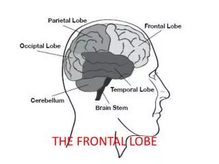
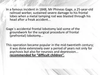
The Frontal Lobes Function and Dysfunction. Frontal lobes: History The frontal lobes as “silent areas”. Frontal lobes: History Phineas Gage (Harlow, 1848). Frontal lobes: History Wartime Trauma.

E N D
Frontal lobes: HistoryWartime Trauma • Improvements in military medicine led to markedly improved survivability from head wounds in WWII and subsequent wars. • Low velocity shrapnel vs. high velocity projectiles. • Example: Luria and localization of function in Russian soldiers.
Frontal lobes: Comparative studiesEvolution • Frontal lobes account for increasing proportion of cerebrum across phylogenetic spectrum (Fuster, 1980) • Prefrontal areas are 33% of cerebral cortical surface in humans (Goldman-Rakic,1984)
Frontal lobes: Comparative studiesWorking memory paradigms • Spatial working memory tasks • Delayed response • Delayed alternation • Feature working memory tasks • Delayed matching to sample • Delayed object alternation
Frontal zones: Primary motor functions • Brodmann area 4 • Input from ventral lateral thalamic nucleus, primary somatosensory area in parietal lobe • Output to internal capsule • Pyramidal motor functions
Frontal zones: Primary motor dysfunction • Initially flacid hemiparesis or hemiplegia on contralateral side • Later spastic hemiparesis or hemiplegia
Frontal zones: Premotor functions • Brodmann area 6 • Input from ventral anterior thalamic nucleus and secondary somatosensory area • Output to motor area and connections via corpus callosum to contralateral premotor area • Integration of sensory and motor information • Praxis
Frontal zones: Premotor dysfunction • Apraxia • Preserved postural praxis via basal ganglia • Contralateral fine motor deficits • Difficulty using sensory feedback
Frontal zones: Frontal eye field functions • Brodmann area 8 • Volitional eye movement in contralateral visual field • Active visual search
Frontal zones: Frontal eye field dysfunction • Failure to move eyes volitionally to contralateral visual field • Intact passive eye movement • Poor visual search
Frontal zones: Dorsolateral functions • Brodmann areas 46, 45,47,8, 9 • Executive functions • Integration of multimodal sensory information • Generation of multiple reponse alternatives • Selection of appropriate response • Maintenance of set, persistence • Set shifting, flexibility • Spatial working memory
Frontal zones: Dorsolateral dysfunction • Difficulty integrating sensory information • Generation of few, stereotyped response alternatives • Poor judgement in response selection • Impersistence • Perseveration
Dorsolateral testsLuria’s reciprocal hand movements & graphic designs
Dorsolateral testsPerseverations within and between Lurian tasks
Frontal lobesPoor organization of learning and recall Copy Free recall
Frontal zones: Orbital functions • Brodman areas 10,11,12,13,14 • Input from limbic and olfactory systems (amygdala, temporal pole, entorhinal cortex, olfactory nerve); inferotemporal lobe areas, ventral visual pathways • Output to autonomic musculature and endocrine system (basal forebrain cholinergic system, caudate, and autonomic system) • Modulation of affective and social behavior; “...preservation of behavioral regulation by external stimuli and its dissolution in the absence of external stimulation.” • Working memory for feature information • Integration of memory and emotional valence • Smell discrimination
Frontal zones: Orbital dysfunction • Disinhibition, socially inappropriate behavior • Failure on feature working memory tasks • Anosmia • Confabulation
Orbital tests • Go-NoGo tasks • UPSIT • Frontal Systems Behavior Scale (FrSBe) • SIGO interview for behavior change
Frontal zones: Cingulate/SMA functions • Brodman areas 24, 32 • Connections with older cortical and deep limbic structures • Drive and motivation • Environmental exploration • Complex attention
Frontal zones: Cingulate/SMA dysfunction • Apathy, akinetic mutism • Alien hand syndrome • Complex attentional deficits, delayed habituation
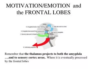
Frontal lobes: Subcortical connections • Parallel but separate connections to caudate and thalamus • Topographic mapping of caudate and thalamus • Subcortical white matter connections • Long tracts • Cortical U-fibers • Small subcortical lesions can mimic large cortical lesions
dorsomedial Frontal lobes: Subcortical connectionsThalamus Dorsolateral prefrontal Anterior Cingulate Orbital prefrontal Dorsolateral caudate Ventromedial caudate Nucleus accumbens Lateral dorsomedial Globus Pallidus Medial Globus pallidus Rostrolateral Globus Pallidus MD Thalamus VA & MD Thalamus VA & MD Thalamus
Frontal lobes: Subcortical connectionsWhite matter cortical u-fibers uncinate fasciculus long tracts to posterior association areas
Frontal lobes: Other signs and syndromes • “Frontal release” signs • Disappear with normal development, reappear in aging or frontal systems dysfunction • Snout, suck, glabellar, grasp • Gegenhalten vs. cogwheeling • Frontal or magnetic gait
Frontal lobes: DementiaKorsakoff’s syndrome This 42 year old man was admitted from a rest home, where he had been displaying a number of problematic behaviors, including compulsive eating of all the vegetables in the garden, and perseverative stuffing of toilets with paper. PMH = Severe alcohol abuse. He had impulsively gone to Florida with a barmaid, and spent all his money. He was “rescued” by his wife, but was unable to return to work as a bartender. He was eventually place in a rest home. NP testing = severe anterograde/retrograde amnesia, moderately severe deficits in executive abilities, marked inertia, abulia, and apathy.
Frontal lobes: TraumaLocalization of TBI from Courville, 1941
Frontal lobes: TraumaContusion with hemorrhage Case history: This 57 year old man was found unconscious by his family, and was thought to have struck his head in a fall. LOC x few hours, but remained confused for the next few days. Sent to a nursing facility, but had repeated arguments with staff, made sexually inappropriate and racist remarks, and was hospitalized. NP testing = severe dysexecutive and disinhibitory deficits, which affected his performance on other tasks.
Dementia of the frontal type vs. Alzheimer’s disease • Preservation of personality & social behavior • Neuropsychological deficits in naming and construction, with frank amnesia after delay • Relative sparing of anterior functions • Generalized and hippocampal atrophy • Early changes in behavior/personality • Neuropsychological deficits in executive and inhibitory functions, no frank amnesia • Relative sparing of posterior functions (must be dissociated from dysexecutive problems) • Anterior atrophy
Frontal lobes:DementiaFrontal type This 56 year old man presented with a one year history of progressive decline in memory and judgment, and several demotions at work. He had displayed a number of socially inappropriate behaviors, such as visiting a neighbor's swimming pool without invitation, neglect of his personal hygiene, and uncharacteristic episodes of anger dyscontrol. NP testing revealed prominent deficits on tests of executive and self-regulatory functions, failures on memory testing typical of frontal impairment (e.g. perseverative and intrusion errors).
Frontal lobes: DementiaFrontotemporal dementia Frontal dementia Normal elderly