Understanding Vestibular System: Anatomy and Physiology Explained
Learn about the vestibular system, its anatomy, function, and common dysfunctions. Dive into the physiology of sensory structures and reflexes involved in maintaining balance.

Understanding Vestibular System: Anatomy and Physiology Explained
E N D
Presentation Transcript
Vestibular Function and Dysfunction Melanie Giesler
Vestibular Dysfunction Prevalence • Four percent of patients18-65 yo visit PCP with complaint of “dizziness” • Three percent consider it “Severely incapacitating” • Third most common complaint in elderly
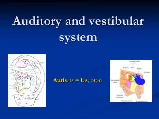
Vestibular System • System of balance • Membranous and bony labyrinth embedded in petrous bone • 5 distinct end organs • 3 semicircular canals: superior, lateral, posterior • 2 otolith organs: utricle and saccule
Vestibular System • Semicircular canals sense angular acceleration • Otolithic organs (utricle and saccule) sense linear acceleration
Vestibular System • Semicircular canals are orthogonal to each other • Lateral canal inclined to 30 degrees • Superior/postereor canals 45 degrees off of sagittal plane
Vestibular Blood Supply • 45% from AICA • 24% superior cerebellar artery • 16% basilar • Two divisions: anterior vestibular and common cochlear artery
Vestibular Nerve Supply • Superior vestibular nerve: superior canal, lateral canal, utricle • Inferior vestibular nerve: posterior canal and saccule
Vestibular Fluid • Membranous labyrinth is surrounded by perilymph • Endolymph fills the vestibular end organs along with the cochlea
Vestibular Fluid • Perilymph • Similar to extracellular fluid • K+=10mEQ, Na+=140mEq/L • Unclear whether this is ultrafiltrate of CSF or blood • Drains via venules and middle ear mucosa
Vestibular Fluid • Endolymph • Similar to intracellular fluid • K+=144mEq/L, Na+=5mEq/L • Produced by marginal cells in stria vascularis from perilymph at the cochlea and from dark cells in the cristae and maculae • Absorbed in endolymphatic sac which connected by endolymphatic, utricular and saccular ducts
Sensory Structures • Ampulla of the semicircular canals • Dilated end of canal • Contains sensory neuroepithelium, cupula, supporting cells
In the Ampulla… • Cupula is gelatinous mass extending across at right angle • Extends completely across, not responsive to gravity • Crista ampullaris is made up of sensory hair cells and supporting cells
In the Ampulla… Hair cells have 50-100 stereocilia and a single kinocilium.
In the Ampulla… stereocilia are not true cilia, they are graded in height with tallest nearest the kinocilium.
In the Ampulla… • Each afferent neuron has a baseline firing rate • Deflection of stereocilia toward kinocilium results in an increase in the firing rate of the afferent neuron • Deflection away causes a decrease in the firing rate
Angular acceleration acts on the hair cells in the Ampulla • Kinocilia are located closest to utricle in lateral canals and are on canalicular side in other canals • Ampullopetal flow (toward the ampulla) excitatory in lateral canals, inhibitory in superior/posterior canals • Ampullofugal flow (away from the ampulla) has opposite effect
Physiology Semicircular Canals • Semicircular canals are paired • Horizontal canals • Right superior/left posterior • Left superior/right posterior • Allow redundant reception of movement • Explains compensation after unilateral vestibular loss
Otolithic Organs • Utricle and saccule sense linear acceleration • Cilia from hair cells are embedded in gelatinous layer • Otoliths or otoconia are on upper surface
Otolithic Organs • Calcium carbonate or calcite • 0.5-30um • Specific gravity of otolithic membrane is 2.71-2.94 • Central region of otolithic membrane is called the striola
Vestibular Physiology • Senses and controls motion • Information is combined with that from visual system and proprioceptive system • Maintains balance and compensates for effects of head motion
Vestibulo-ocular reflex • Vestibulo-ocular reflex • Membranous labyrinth moves with head motion • Endolymph does not causing relative motion • Cupula on right canal deflected towards utricle causing increase in firing rate, left deflects away causing a decrease in firing rate. • Reflex causes movement of eyes to the left with saccades to right • Stabilizes visual image
Nystagmus • If acceleration stops, and spin to right is at constant velocity, sensation of motion stops after 14-20 seconds as does nystagmus • Cupula only takes 8-10 seconds to return to equilibrium position • Vestibular integrator is the term for the prolongation and is mediated by the vestibular nuclei and cerebellum
Vestibulospinal Reflex • Senses head movement and head relative to gravity • Projects to antigravity muscles via 3 major pathways: • Lateral vestibulospinal tract • Medial vestibulospinal tract • Reticulospinal tract
Office Examination of the Dizzy Patient • Dix-Hallpike Maneuver • Used to provoke nystagmus and vertigo commonly associated with BPPV • Head turned 45 degrees to maximally stimulate posterior semicircular canal • Head supported and rapidly placed into head hanging position • Frenzel glasses eliminate visual fixation suppression of response
Dix-Hallpike Manuveur • Positive test • Up-beating nystagmus • Nystagmus to the stimulated side • Rotary component to the affected ear • Lasts 15-45 seconds • Latency of 2-15 seconds • Fatigues easily
Pneumatic Otoscopy • Positive and negative pressure applied to middle ear • Hennebert’s sign/symptom – nystagmus and vertigo with pressure, alternates with positive and negative pressure • Can be present in patients with perilymphatic fistula, syphilis, Meninere’s disease, SCC dehiscence syndrome
Head Shake Nystagmus • Evaluates unilateral vestibular weakness • Head tilted back 30 degrees • Shake back and forth for 30 seconds as quickly as possible • Unilateral vestibular deficit causes slow phase nystagmus to the side of lesion • Low sensitivity (27%) • Good specificity (85%)
Head Thrust Test • Inhibitory response not as robust as the stimulatory response to stimulate VOR • Movements that overcome the inhibitory response of vestibule will result in VOR lag • Head tilted 30 degrees • Rapid head movements to either side with focus on examiner’s nose • Patients have catch-up saccade when rotated to side of weakness • Sensitivity 75%, Specificity of 85%
Dynamic Visual Acuity • Used for bilateral vestibular weakness • Visual acuity checked on Snellen chart • Rechecked while rotating head back and forth at 1-2 Hz. • Loss of 2-3 lines considered abnormal – usually b/l loss due to ototoxicity or aging.
Romberg Test • Patient asked to stand with feet together and eyes closed • Fall or step is positive test • Equal sway with eyes open and closed suggests proprioceptive or cerebellar site • More sway with eyes closed suggests vestibular weakness
Fukuda Stepping Test • Originally described by Fukuda using 100 steps on a marked floor. • Patients are asked to step with eyes closed and hands out in front • Rotation by more than 45 degrees is abnormal • Rotation usually occurs to the side of the lesion • Rotation often found in asymptomatic patients
Dysdiadochokinesia Testing • Most commonly tested with the hand slapping test • Abnormalities seen in patients with cerebellar dysfunction • Poor sensitivity and specificity
Tandem Gait Test • Patients are asked to walk heal to toe in a straight line or in a circle • Complex function evaluates many aspects of balance • Poor performance seen in cerebellar lesions, but can be seen in many disorders • Poor sensitivity and specificity
Orthostatic Hypotension • Most often in patients on BP meds with “light headedness” on sitting to standing • Defined as drop of SBP 20mm HG or DPB 10mm HG within 3 minutes of standing • Tilt exams offer objective measurements with well established norms • Patients with no symptoms will often “Tilt”
Voluntary Hyperventilation • Patients asked to over breathe for 90 seconds to 3 minutes (elevate pH – vasoconstriction) • Hyperventilation causes symptoms in some anxiety disorders • May provoke symptoms in normal • Poor test for vestibular diagnosis but can elevate pH, increase 8th nerve firing - lesions in petrous apex, acoustic schwannoma, 8th nerve demylination
Electronystagmography (ENG) • Divided into oculomotor tests, positional and positioning tests, and caloric tests • Only vestibular test with the ability to test individual labyrinths separately • Relies on the vestibulo-ocular reflex (VOR) to test the peripheral vestibular function • Mostly a test of HSCC function
Electronystagmography (ENG) • Oculomotor tests • All test eye movements that originate in the cerebellum • Saccadic tracking • Smooth pursuit tracking • Optokinetic testing
Oculomotor Tests • Saccadic tracking • Patients concentrates on a randomly moving target • Latency – difference in time between movement of object and eye (150-250 ms) • Velocity – speed of saccade 200-400 degrees/second low end of normal • Accuracy – amount of undershoot/overshoot of target (75-120%)
Smooth Pursuit Test • Tests ability to accurately and smoothly pursue a target • Gain of eyes compared to movement of target • Saccade movements eliminated from calculations • Asymmetrical pursuit highly suggestive of central disorders
Optokinetic Tests • Vestibular system and optokinetic nystagmus allow steady focus on objects • Target is rapidly passed in front of subject in one direction, then the other • Eye movements are recorded and compared in each direction • Asymmetry suggestive of CNS lesion • High rate of false positive results
Caloric Testing • Established and widely accepted method of vestibular testing • Most sensitive test of unilateral vestibular weakness • Patient positioned 30 degrees from prone (HSCC vertical allowing max stim) • Cold and warm water/air flushed into EAC