Download

1 / 24
270 likes | 629 Views
Effects of PPV on the Pulmonary System. Chapter 17. Pulmonary Complications. Lung Injury Gas distribution Pulmonary blood flow VAP Hypoventilation Hyperventilation Air trapping Oxygen toxicity ↑ WOB Patient-Ventilator dyssynchrony Mechanical problems

E N D
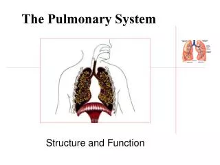
Effects of PPV on the Pulmonary System Chapter 17
Pulmonary Complications • Lung Injury • Gas distribution • Pulmonary blood flow • VAP • Hypoventilation • Hyperventilation • Air trapping • Oxygen toxicity • ↑ WOB • Patient-Ventilator dyssynchrony • Mechanical problems • Complications of the artificial airway
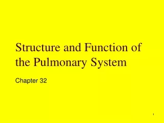
Bronchi fan out like coral in this resin cast that also shows pulmonary arteries and trachea. The bronchi supply air and pulmonary arteries supply blood to the lungs. Together they take in air from the atmosphere, oxygenate the blood, and excrete the carbon dioxide back out of the body. Photograph by Martin Dohrn/Royal College of Surgeons/Science Photo Libraryhttp://science.nationalgeographic.com/science/photos/lungs/lungs-cast.html
VALI: lung injury as a consequence of mechanical ventilation VAP Air trapping Patient-ventilator dyssynchrony Extra-alveolar gas VILI: occurs at the level of the acinus, microscopic level of injury Biotrauma Shear stress Surfactant depletion Lung Injury
Barotrauma • Trauma associated with pressure • Can result in the formation of extra-alveolar gas • Predisposed to developing with: • High peak pressures with low end-expiratory pressures • Bullous lung disease • High PEEP with high Vt • Aspiration of gastric contents • Necrotizing pneumonias • ALI/ARDS • Gas under pressure causes alveolar rupture
Subcutaneous emphysema Puffing in the skin Crepitant Usually occurs without complication Pneumomediastinum May lead to compression of esophagus, great vessels, and heart Treatment depends on severity i.e. cardiac tamponade Pneumothorax Lung collapse on affected side Shift of mediastinum away from affected side Resonant/hyperresonant percussion Treat with a chest tube Pneumoperitoneum Generally follows pneumomediastinum Air dissects into the retroperitoneal space Can interfere with the movement of the diaphragm Barotrauma
The peak pressure alarm is activated on a ventilated patient. Assessment of the patient reveals puffing of the skin of the patient’s neck and face, which feels crepitant to the touch. The right hemithorax is hyperresonant to percussion and breath sounds are absent. What would be an appropriate action for the RT? Physical finding indicate the presence of a right-sided pneumothorax. A physician should be contacted for an order for a CXR and to begin treatment. The RT should stay with the patient and make sure the pneumothorax does not become a tension pneumothorax. Appropriate emergency equipment should be kept close at hand, may need to manually ventilate until treatment can be administered. Clinical Rounds 17-1, p.359
Volutrauma • Increasing volume overdistends areas of the lungs • Associated with iatrogenic lung injury • Due to regional differences in lung compliance, PPV tends to produce larger volumes in more compliant areas • Causes biotrauma
Atelectrauma • Underinflation of the lung units • Injuries that occur because of repeated opening and closing of lung units at lower lung volumes • Three primary types • Shear stress • Alteration and washout of surfactant • Microvascular injury • Described as alveolar rupture, interstitial emphysema or perivascular and alveolar hemorrhage →death
Biotrauma • Mechanical stress disrupts normal cell function • Strains normal cell configuration • Inflammatory response in the lungs • Cytokines • Tumor necrosing factor • Damage from ventilator mismanagement can be indistinguishable from ARDS
Multiple Organ Dysfunction: chemical mediators can leak into the blood vessels leading to inflammatory responses in the liver, gut, and kidneys • Vascular endothelial injury: pressure changes pull fluid into the interstitial space = edema • One of the first studies to demonstrate this and recommend lung protection strategies was in 1970!
Two days after admission to the hospital a patient with acute pancreatitis requires mechanical ventilation. Although ventilation is well maintained with ventilator oxygenation becomes a problem. The PaO2 is 70mmHg on 75%. The patient is on PCV with a set pressure of 20cmH2O and a current PEEP of 5cmH2O. Auscultation reveals bibasilar crackles and scattered crackles in the posterior basal segments. What change in therapy might be appropriate? The crackles in the basilar and posterior areas may indicate atelectasis and the opening and closing of alveoli in dependent areas. An increase in PEEP is indicated and a recruitment maneuver might also be considered. Clinical Rounds 17-2, p. 363
Gas Distribution & Pulmonary Blood Flow • Spontaneous breathing favors gas distribution to the dependent lung areas and periphery • PPV impacts dead space • Normal pulmonary blood flow favors gravity dependent areas and central areas of the lungs • PPV can affect PVR
VAP Pneumonia acquired >48hours after intubation Rates are increased by Invasive catheters and monitoring devices Predisposing illnesses or disorders Injury to nasopharynx or tracheal surface Decreased effectiveness of cough Bypassing upper airway defense mechanisms Reduced healing if the nutritional status is poor Diagnosis Fever > 38.2°C ↑ WBC Purulent secretions/aspirate New infiltrates on CXR Causes Chronic microaspiration of subglottic secretions Nosocomial Infections
Prevention of VAP • Handwashing • Oropharyngeal cleaning/decontamination • Noninvasive ventilation • HOB>30° • Kinetic beds • Stress ulcer prophylaxis • Selective digestive tract decontamination • Care of ETT or tracheostomy tube • CASS • Ventilator circuit management • Prophylactic antibiotics • Infection control to monitor
Four days after intubation and mechanical ventilation a 68 y.o. patient has the following findings: fever of 39°C, WBC count of 18,000 cells/ml, and a recently developed LLL infiltrate. Secretions are thick and yellow to green in color. What therapeutic interventions might benefit this patient? These findings are consistent with VAP. Collection of a sputum sample (with bronchoscopy) to identify the causative organism and direct antibiotic therapy. Consider VAP prevention strategies Clinical Rounds 17-3, p. 370
Acid-Base Status • Hypoventilation • Hyperventilation • Metabolic acid-base imbalances
A patient has been mechanically ventilated for 7 days. The patient’s normal baseline ABG’s on RA are 7.38/51/58/29. Current ABG’s on VC-SIMV 8, Vt 800ml, FiO2 .25 are 7.41/40/67/24. The patient is not spontaneously breathing. The VC-SIMV mandatory rate is reduced to 4. The patient's spontaneous rate increases to 28 spont Vt is 250ml, SpO2 drops from 95% to 91%. The patient appears anxious. What is the problem? The ABG values after 7 days of PPV are normal. However this patient’s baseline suggest chronic CO2 retention. The patient has been hyperventilated with the ventilator and the kidneys have reduced the HCO3 level to normal. When the mandatory rate is reduced for weaning, the PaCO2 rises, stimulating spontaneous ventilation. Unfortunately this patient cannot maintain a normal PaCO2 and pH as suggested by the high spontaneous rate. To correct the problem the patient’s mandatory rate must be reduces gradually until normal baseline ABG values are restored. Provide appropriate PSV for spontaneous breaths. Clinical Rounds 17-4, p. 372
Auto-PEEP • Unintentional PEEP that occurs with patient receiving ventilatory support when a new inspiratory breath is begun before expiratory flow has ended
Auto-PEEP • Occurs in three distinct forms: • Active contraction of expiratory muscles during exhalation • Presence of high Ve, short Te, and increased expiratory resistance • Airflow obstruction/airway collapse • Affects ventilator function • Reduce auto-PEEP by: • Higher inspiratory gas flow • hypoventilation
A patient with COPD is receiving VC-CMV mode. The set Vt is increased from 700 to 900ml and the rate is increased from 10 to 18. The RT notices a progressive rise in PIP. Vt are transiently less than 850ml after the change. Eventually the exhaled Vt reads 850ml. Baseline pressures remain at 0. The patient appears unable to trigger a breath and is using accessory muscles to trigger the breath. What is the most likely cause of this problem? The new Ve is 16.2L/min. The increase in Ve resulted in auto-PEEP which caused the rise in PIP and the transient drop in exhaled Vt that occurred after the change. Also, the patient is unable to trigger the ventilator another possible indication of air trapping Clinical Rounds 17-4, p.375
Hazards of Oxygen Therapy • Oxygen toxicity • Absorption atelectasis • Depression of ventilation
System imposed WOB VC-SIMV with PSV has the greatest WOB WOB during weaning Decreased support from ventilator Reducing WOB Artificial airway Setting machine sensitivity and inspiratory flow Patient ventilator synchrony Reducing Ve demands WOB
Mechanical and Operational Hazards • Ventilators are SAFE when monitored with care • Cause of problems comes from staffing, communication, training • Correct alarm settings are critical • Artificial airway complications