Download

1 / 17
180 likes | 331 Views
Learn about Peyronie’s disease, its diagnosis through ultrasound, X-ray, MRI, and treatments such as penile fracture management and collagen fibrosis prevention. Explore calcified plaques, fibrosis, and more.

E N D
X- ray pelvis showing bilateral fibrosis of the corpora cavernosa tunica albuginea denoting peyronie’s disease ( yellow line)
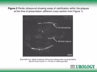
Calcified plaque involving right corpus cavernosum ( painful erection) ( Peyronie’s disease)
Collagen in peyronie’s disease. Densely packed collagen fibers prevent normal sliding of tissue during expansion & contraction of erection process resulting in a curved penis. Collagen in normal tissue of tunica albuginea. Organized collagen fibers arranged in sheets that slide past each other during expansion & contraction of the erection process.
H. & E. showing fibrosis of tunica albuginea but does not affect corpora cavernosa . It may be calcified or ossified (os penis)
Mallory stain showing fibrosis of tunica albuginea in a caseof peyronie’s disease
Dorsal neurovascular bundle is lifted off the underlying corporal body by a blue rubber loop. So nerve function will be preserved. The actual problem is the corporal body where incision & grafting will take place.
Penile fracture (Broken penis)It affects directly the tunica albuginea. Dignosis is by cavernosography ; MRI & urethrogrphy to rule out whether the urethra is traumatized, No bone inside cavernous tissue.