Download
1 / 93
930 likes | 1.11k Views
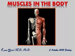
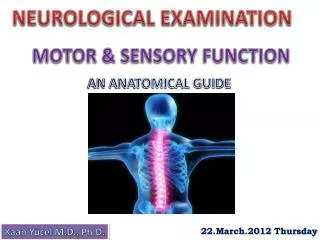
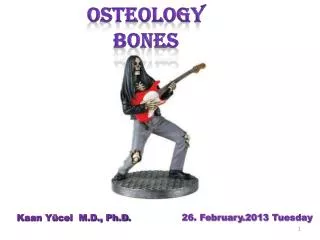
OSTEOLOGY BONES. 26. February . 201 3 Tuesday. Kaan Yücel M.D., Ph.D . . INTRODUCTION TO OSTEOLOGY. Osteology ( Gk , osteon, bone, logos, science) branch of medicine concerned with the development and diseases of bone tissue The human skeleton 206 bones in adults .

E N D
OSTEOLOGY BONES 26. February.2013 Tuesday Kaan Yücel M.D., Ph.D.
.INTRODUCTION TO OSTEOLOGY Osteology (Gk, osteon, bone, logos, science) branch of medicine concerned with the development and diseases of bone tissue The human skeleton 206 bones in adults
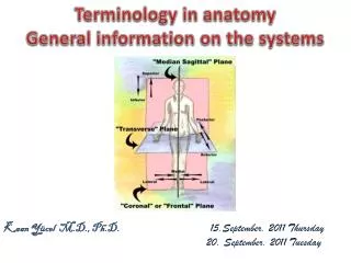
The skeletal system may be divided into • 2functional parts: • The axial skeleton • head (cranium or skull) • neck (hyoid bone and cervical vertebrae) • trunk (ribs, sternum, vertebrae, and sacrum) • The appendicular skeleton • Limbs • including those forming the shoulde & pelvic girdles
Bone one of the hardest structures of the animal body calcification of its extracellular matrix some elasticity results from the organic matter great rigidity results from their lamellous structures and tubes of inorganic calcium phosphate colorin a fresh state pinkish-whiteexternally, deep red within.
HISTOLOGY OF THE BONE sparse cells surrounded by an extracellular network/matrix
Osteoblasts secrete proteins into the matrix.
Mature bone is composed of proteins and minerals. 60% the weight of the bone mineral Rest- water & matrix. 90% of the matrix proteins collagen1/3 of the bone weight very strong forms bone, cartilage, skin, and tendons. High resolution image of cortical bone and single collagen fibril (inset)
Mineralsof the matrix Mainlycalcium phosphate & calcium carbonate Embedded in the protein network Providehardness and compressive strength.
Matrix maintained by osteocytes Haversiansystems or osteons concentric rings of osteocytes arranged around a central blood vessel.
Principal types of bone cells Osteogenic cells Osteoblasts Osteocytes Osteoclasts
Periosteum membranesurroundingthe bone tissue provides a routeforthevasculatureandnervesupply. participates in bone growthandrepair. Endosteum linesthe marrow cavity active during bone growth, repair, and remodeling covers trabeculaeof spongy bone lines the inner surfaces of the central canals
CartilageSand Bones • The skeleton is composed of cartilages and bones. • Cartilage • resilient, semirigid form of connective tissue • forms parts of the skeleton where more flexibility is required. articulating of bones participating in a synovial joint capped with articular cartilage provides smooth, low-friction, gliding surfaces for free movement
Blood vessels do not enter cartilage avascular • Diffusion • bone /cartilage in the skeleton • changes as the body grows • younger a person the more cartilage • bones of a newborn are soft and flexible because mostly composed of cartilage.
CartilageSand Bones The skeleton is composed of cartilages and bones. The amount and kind of extracellular fibers in the matrix depends on the type of cartilage. Heavyweightbearing areas or areas prone to pulling forces Morecollagenfibers, lessflexiblecartilage.
Functionsof cartilage support soft tissues provide a smooth, gliding surface for bone articulations at joints enable the development and growth of long bones.
Typesof cartilage • 1. Hyaline • most common,matrix w/ moderate amount of collagen fibers articularsurfaces of bones • 2. Elastic • large number of elastic fibers external ear • 3. Fibrocartilage • limited number of cells &ground substance amidst substantial amount of collagen fibers intervertebral discs
Bones function as • supportive structures for the body • protectors of vital organs • reservoirs of calcium and phosphorus • levers on which muscles act to produce movement • containers for blood-producing cells
TYPES OF BONES • according to their shape gross anatomy • Long bones • tubular humerusin the arm • 3)Flat bones • protectivefunctions • flat bones of the cranium protect the brain • 2)Short bones • cuboidal • tarsus (ankle) carpus (wrist)
Classification of Bones • 4) Irregular bones • various shapes other than long, short, or flat • bones of the face
Classification of Bones • 5) Sesamoidbones • patella or knee cap • protect the tendons from excessive wear • often change the angle of the tendons as they pass to their attachments.
Long bones develop by replacement of hyaline cartilage plate endochondral ossification a shaft diaphysis - two ends epiphyses Metaphysis a part of the diaphysis adjacent to the epiphyses. Diaphysisencloses the marrow cavity.
2types of bones according to histological features compact bone &spongy (trabecular) bone relative amount of solid matter #&size of the spaces they contain
All bones have a superficial thin layer of compact bone • around a central mass of spongy bone • except where the spongy boneis replaced by a medullary (marrow) cavity. • Spongy bone • found @ expanded heads of long bones +fills most irregular bones. • Compact bone • forms outer shell of all bones+shafts in long bones.
Bone Markings and Formations • Bone markings appear wherever tendons, ligaments, and fascias are attached or where arteries lie adjacent to or enter bones. • Other formations occur in relation to the passage of a tendon (often to direct the tendon or improve its leverage) or to control the type of movement occurring at a joint.
Bone Markings and Formations • Surfaces of the bones are not smooth. • Bones display elevations, depressions and holes. • The surface features on the bones are given names to distinguish and define them.
Vasculature and Innervation of Bones • Bones are richly supplied with blood vessels. • Veins accompany arteries. • Nerves accompany blood vessels supplying bones.
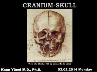
Skull is supported on the summit of the vertebral column, and is of an oval shape, wider behind than in front. It is composed of a series of flattened or irregular bones which, with one exception (the mandible), are immovably jointed together. It is divisible into two parts: cranium, which lodges and protects the brain, consists of 8 bones skeleton of the face,of 14
Occipital bone at the back and lower part of the cranium trapezoid in shape and curved on itself. pierced by a large oval aperture, the foramen magnum, cranial cavity communicates with the vertebral canal throughtheforamenmagnum
Parietal Bones form, by their union, the sides and roof of the cranium each bone irregularly quadrilateral in form external surface convex, smooth
Frontal Bone @front of the skull. Forms theforehead. Entersinto the formation of the roofs of the orbital and nasal cavities.
Temporal Bones at the sides and base of the skull. consist of the pathway to the inner ear and contributes to the formation of the jaw with the mandible.
Sphenoid Bone at the base of the skull in front of the temporals and basilar part of the occipital. median portion or body, two great and two small wings extending outward from the sides of the body, and two pterygoid processes which project from it below. supplies the bed for the pituitary gland.
Ethmoid bone exceedingly light and spongy cubical in shape at the anterior part of the base of the cranium between the two orbits, at the roof of the nose contributes to each of these cavities.
Cranial Fossae Anterior cranial fossa occupiedbytheinferior and anterior parts of the frontal lobes of the brain shallowest cranial fossa Middle cranial fossa butterfly-shaped central part composed of the sellaturcica on the body of the sphenoid large, depressed lateral parts on each side Posterior cranial fossa largest and deepest cranial fossa formed mostly by the occipital bone
Facial Bones • Nasal Bones • two small oblong bones, varying in size form in different individuals • placed side by side @ middle &upper part of the face • form, by their junction, “the bridge” of the nose. • Maxillæ(Upper Jaw) • largest bones of the face, excepting mandible • form the whole of the upper jaw. • Formthe boundaries of 3 cavities • roof of the mouth • floor and lateral wall of the nose • floor of the orbit
Facial Bones Lacrimal Bone smallest & most fragile bone of the face at the front part of the medial wall of the orbit ZygomaticBone (Malar Bone) small and quadrangular at the upper and lateral part of the face forms prominence of the cheek part of the lateral wall & floor of the orbit. Zygomaticarch zygomaticprocess of the temporal bone temporal process of the zygomaticbone
Facial Bones Palatine Bone @ back part of the nasal cavity. contributes to the walls of three cavities floor and lateral wall of the nasal cavity roof of the mouth floor of the orbit. Inferior Nasal Concha extends horizontally along the lateral wall of the nasal cavity.
Facial Bones Vomer in the median plane thin, somewhat quadrilateral in shape forms hinder & lower part of the nasal septum. Mandible (Lower Jaw) largest and strongest bone of the face serves for the reception of the lower teeth.
Facial Bones • Hyoid Bone • shaped like a horseshoe • suspended from the tips of the styloid processes of the temporal bones.
Ribs (L. costae) • curved flat bones • form most of the thoracic cage. • 3 types of ribs: • True (vertebrocostal) ribs (1st-7th ribs): • directly to the sternum. • False (vertebrochondral) ribs • (8th, 9th, and usually 10th ribs): • indirect withthesternum • Floating (vertebral, free) ribs • (11th, 12th, and sometimes 10th ribs): • No connectionwiththesternum
Typical ribs (3rd-9th) have the following components: • Head • one facet for articulation with the numerically corresponding vertebra • one facet for the vertebra superior to it • Neck • Tuberclearticulates with the corresponding transverse process of the vertebra. • Body (shaft) • .
Costal cartilages prolong the ribs anteriorly and contribute to the elasticity of the thoracic wall, providing a flexible attachment • for their anterior ends.
Intercostal spaces separate the ribs and their costal cartilages from one another. • The spaces are named according to the rib forming the superior border of the space—for example, the 4th intercostal space lies between ribs 4 and 5. • There are 11 intercostal spaces and 11 intercostal nerves. Intercostal spaces are occupied by intercostal muscles and membranes, and two sets (main and collateral) of intercostal blood vessels and nerves, identified by the same number assigned to the space.